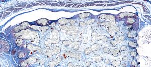

Within hematology research, I’m interested in analyzing cytological smears. Through blood smears, I look at individual cells and their morphology. It is necessary to analyze the subcellular structures to quantify the number of cells and morphology.
Challenges to be solved:
Without the O8, it would be necessary to analyze a sample manually with a traditional microscope without much flexibility. We might be able to take a few individual pictures from individual views. This would take a lot of time. Hence, the O8 cannot really be compared to this workflow, as it simply offers many new opportunities. When comparing the O8 to other scanners, the resulting image quality definitely makes a key difference. Good focusing is key since oil immersion has a shallow focus depth. With the O8, I can set focus points resulting in excellent images. These specific parameters (oil immersion in high resolution) are exceedingly rare to find on the market.
Only very few companies and products on the market offer oil scanning capability. We have seen a couple of scans from other market participants but were not fully convinced. We need to see subcellular structures, and analyzing the chromatin, nuclear shape, cytoplasmic structures, etc. is key for us, which is why we need the best possible resolution for digital images. In conclusion, the capability to scan with both 60x and 100x oil immersion microscopy is a niche that PreciPoint is very strong in and the image quality of the O8 convinced us.
Stay Ahead with Insights from Precipoint!
Welcome to our newsletter! Be the first to know about our latest products, services, webinars, and happenings in PreciPoint. Don't miss out on this opportunity to stay informed. Subscribe to our newsletter today!
By clicking “Subscribe”, you agree to our privacy policy.
“It is intuitive. It offers high-quality images. It is a hybrid between a microscope and a scanner, offering many new and interesting possibilities.”

Our approach and solution:
The main advantage to have samples in electronic form is that it opens completely new possibilities that have not been available up until now. I can for example, analyze the digital slides with analysis software. I can also share them much more easily with other colleagues, compared to having a physical slide. I can show or send them the digitized scan to analyze for consultation. Before, it would be necessary to analyze a sample manually using a traditional microscope without much flexibility.
The O8 is extremely easy to use and it did not take a lot of time to get used to the operations. It is extremely helpful for my project since it is very intuitive, and you do not deal with a huge overhead of technology controls that you have to learn. It is incredibly attractive to look at things you are used to seeing in a conventional microscope and seeing it on a computer. On top of that, I also find it much more comfortable that I do not have to look through oculars.