
In our virtual discussion with Science Manager Paul Jank „How Can Digital Microscopy Accelerate Your Research Work? “, we looked into recent digital research works at the Institute for Pathology at the Philipps University in Marburg.
In this article, we give an overview of the opportunities of working digitally in research today and bring some key considerations to help research institutes get started and get the most out of their digitization efforts.
Digital Microscopy Advances Research Collaboration
Paul Jank has been working with digital microscopes and slide scanning for the past 6 years. This has made his work much more integrative. Digitization has accelerated internal processes and facilitated national and international collaboration. Paul explains how digitized slides have improved his daily work:
• Non-physician personnel are included in the scanning workflow.
• Oversea partners examine scans simultaneously.
• Cooperation partners analyze the same image and can exchange on the exact same basis.
• The digitized specimens are quickly accessed and shared.
Slides are available instantly
In a nutshell, Paul considers the speed with which he can access and analyze digitized slides to be the most compelling benefit of working digitally. After scanning, the Whole Slide Images (WSI) are available at any time and from anywhere.
Digitization has drastically cut the time between sample preparation and analysis. The research personnel no longer need to look through the physical archive for the samples or even mail them to the cooperation partner. In a structured digital file system, you can retrieve the scans easily and share them immediately.
Digital Images Key Enabler For Artificial Intelligence
Digitization opens new avenues in many areas – Research is no exception: Whole Slide Images have made the use artificial intelligence (AI) algorithms in histology research projects possible.
The Institute for Pathology in Marburg is riding the AI wave and is already conducting research projects digitally. With scans minimizing interobserver-variability, the field of biomarker validation is a very good example of how working digitally can improve research outcomes.
Stay Ahead with Insights from Precipoint!
Welcome to our newsletter! Be the first to know about our latest products, services, webinars, and happenings in PreciPoint. Don't miss out on this opportunity to stay informed. Subscribe to our newsletter today!
By clicking “Subscribe”, you agree to our privacy policy.

In tissue research, every PhD student at the institute is now working with artificial intelligence and are writing validated syntaxes in order to run evaluation schemes with AI in the future."
Paul Jank, Science Manager and Biotechnologist at the Institute of Pathology, Philipps University of Marburg.
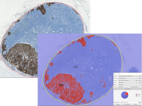
Biomarker Evaluation with Tissue Micro Arrays
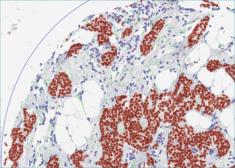
In the research field, the Institute for Pathology has made a particularly successful use of artificial intelligence with the Tissue Micro Array (TMA) method. In order to standardize the evaluation of immunohisto-biomarkers, the AI was trained to automatically distinguish tumor cells from stroma and to color-code lymphocytes. Thus, an immunohistological evaluation can be made within minutes for approximately 1000 different tissue sections. The data can then be used very easily for statistics.
Compared to analog processes, the new method opens new possibilities and allows for a significant performance increase. Manually, the researcher would have to evaluate each sample individually. Without TMA, each of the 1000 slides would first have to be stained.
AI-Enabled Research and Computational Pathology
Currently, the institute is working intensively with automatic image analysis, so-called computational pathology. In a current research project, lymph nodes are stained with pancytokeratin to determine the percentage of infiltration of tumor cells. With the digital slides, the percentage of tumor cells vs. healthy cells and the infiltrated areas in the lymph node can be evaluated much more easily and precisely.
Students benefit from digitization
The Institute for Pathology at the University of Marburg now also works digitally in the area of teaching. The images generated with the digital microscope are integrated into a cloud-based learning platform and made available to students in medicine and human biology for online microscopy. The platform eliminates the need for on-site microscopy, which has become critical during corona times, and allows students to study remotely for exams through slide annotations and supplemental material.
Technical Considerations For Scanning
250,000: That’s the number of samples Paul wants to digitize retrospectively at the Marburg Institute for research purposes and for the German Breast Group’s biobank of clinical breast cancer studies. Around 50 to 100 new research samples come in every day. Currently, the institute has a digitization throughput of around 350 slides per day. This requires a suitable storage solution and infrastructure, considering that each individual digitized slide has an average file size of 500 to 1000 MB.
There are indispensable solutions for reducing the file size during scanning, such as good file compression. But tissue recognition also provides good results. This analysis detects the tissue to be scanned on the slide and omits the background and gel during scanning. This results in smaller image files, which directly reduces the amount of storage space required. Learn more about tissue recognition here.
Getting started with Digitization
Before you start digitizing, you should clearly define your long-term goals. However, digitization should always aim to make the workflow more efficient. Don’t digitize everything at once. A step-by-step approach is preferable.
Therefore, consider in advance exactly at which points of the workflow digitization makes the most sense and will bring you early results. If possible, start where you can avoid any interruption of the workflow and where digitization will accelerate the process.
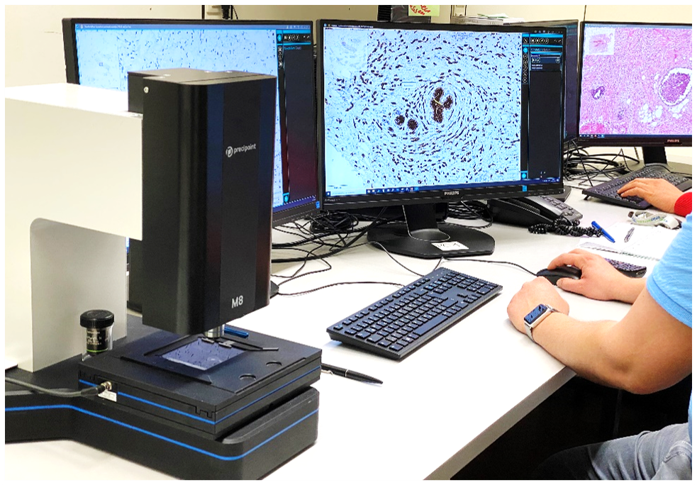
Paul has been using the M8 microscope and scanner to digitize samples for a few years. The compact device offers him flexibility in his work.
“The device has intuitive operation and is installed within a few minutes. The PreciPoint image format vmic is widely applicable. And if something does go wrong, the customer service team is quick to help,” concludes Paul.
Are you interested in digital tissue research?
Discover our Microscope and Slide Scanner M8, our all-in-one device for digital microscopy and slide scanning needs.