
Virtual microscopy is widely used in contemporary pathology education and assessment as it facilitates efficient analysis, diagnosis, and annotation. Educators, students, and other medical professionals achieve high-quality results quickly as compared to when using traditional microscopy. Because of its many advantages, virtual microscopy is popular among students and educators worldwide. Today we will discuss the application of virtual microscopy in learning and assessment and talk about some of the most important advantages of using virtual microscopy in education or research work.
Virtual microscopy is a technology that allows educators and students to access high-resolution digital images of microscope slides, enabling them to explore and analyze specimens in ways that were previously impossible. This powerful tool is used in teaching to enhance the learning experience of students in various fields such as medicine, biology, and pathology and has opened new opportunities for them. With the help of high-quality software and secure cloud platforms to share, annotate, and evaluate virtual slides, students and researchers can now do complex tasks more easily than before.
Stay Ahead with Insights from Precipoint!
Welcome to our newsletter! Be the first to know about our latest products, services, webinars, and happenings in PreciPoint. Don't miss out on this opportunity to stay informed. Subscribe to our newsletter today!
By clicking “Subscribe”, you agree to our privacy policy.
Ease of Accessibility
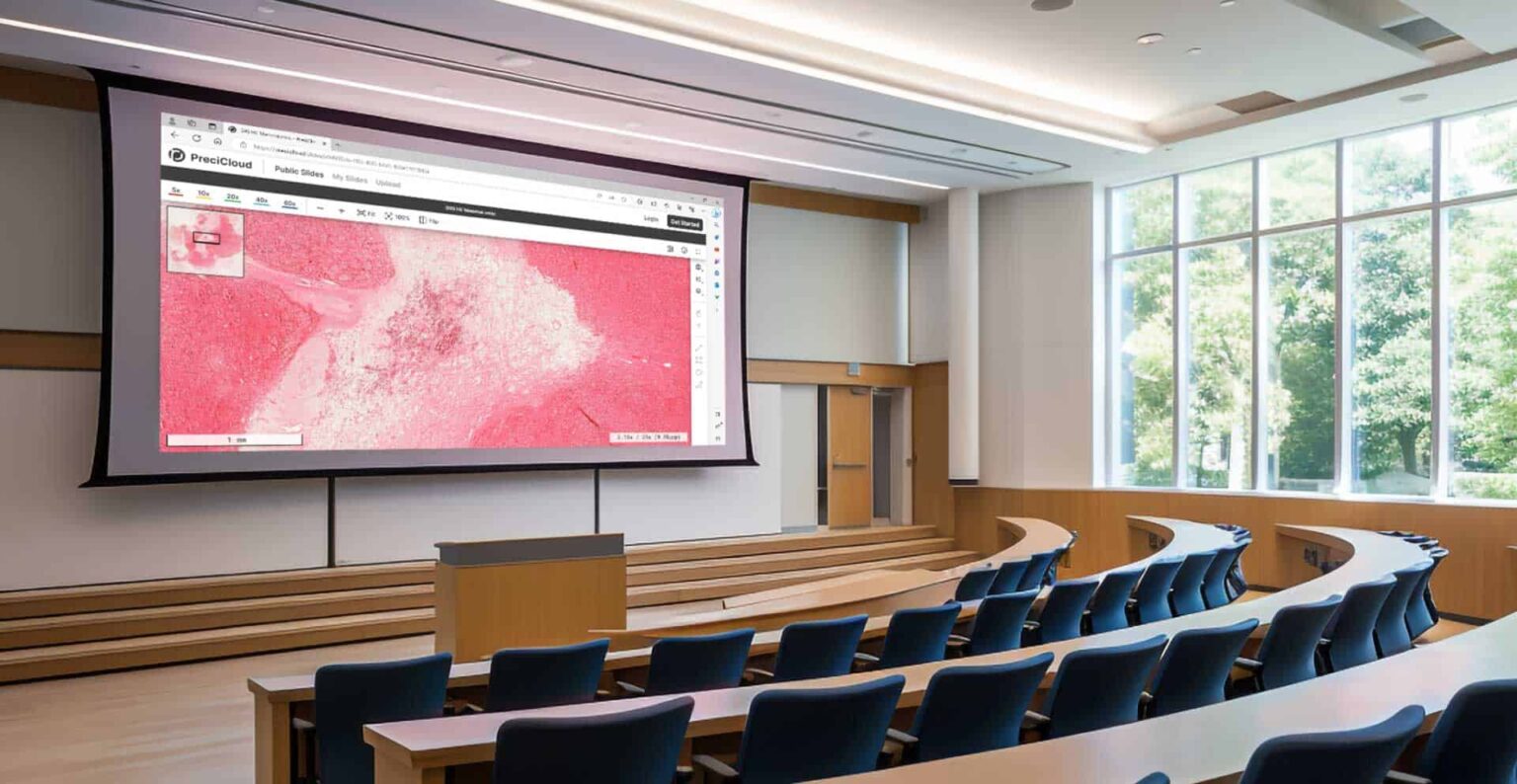
Virtual microscopy has a distinct advantage in terms of accessibility. Students can access digital image slides from anywhere with an internet connection, allowing them to engage in their sessions with ease. It benefits learners scattered worldwide, as it enables them to access top-quality educational resources from around the world. Thus, virtual microscopy has emerged as an indispensable tool in facilitating accessible and effective education.

Virtual microscopy offers two additional critical benefits, namely cost-effectiveness and work efficiency. When it comes to traditional methods of learning pathology, educators and learners need a certain amount of time to prepare and maintain the slides. These slides are then required to be transported to certain locations where the learners and the educators observe them. That makes the whole process quite complicated and time-consuming. On the contrary, virtual microscopy decreases the time needed for preparation, maintenance, and transportation making the process faster, easier, and more efficient.
Unique Learning Experience
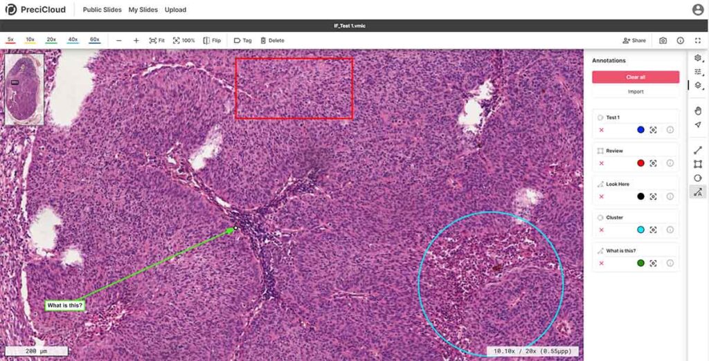
When applying virtual microscopy in contemporary pathology classes, students can have a unique learning experience. The experience is distinct in nature as it encompasses not only the distribution of knowledge but also involves interactive activities between the participants of the session. This kind of learning experience improves knowledge retention power, which students can easily apply later in their professional life; the learning becomes more inclusive. Some of the tasks that students can undertake using virtual microscopy are:
- Zoom in and zoom out of images.
- Annotate and mark the areas of interest.
- Compare diverse slides slide-by-slide.
Virtual microscopy can also be used to create simulations that allow students to practice diagnosis and treatment in a safe and controlled environment. With such detailed knowledge and practical experience, the learning becomes more engaging and comprehensive, which affects the results and learning outcomes.
Possibilities for Learning
Educators can create customized learning materials for their students. They can create a collection of slides that can be tailored to certain learning objectives and purposes, allowing the students to practice analysis and diagnostics on these samples. This exposes learners to a wide range of pathological specimens that are relevant to their area of study.
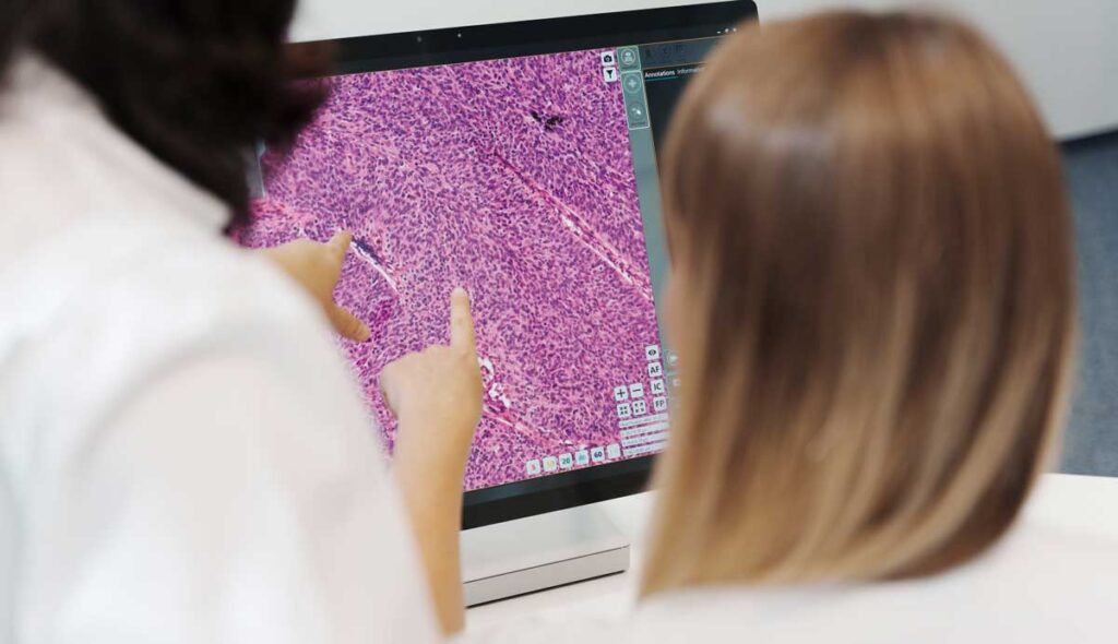
Collaborative learning is another important advantage of virtual microscopy. Students can share their findings and annotations with co-learners. That can facilitate discussion and peer review. As a result, active learning is enabled as students get engaged to actively manage the material and apply their knowledge to real-world scenarios.
Virtual Microscopy in Assessment
Virtual microscopy is being actively used for the assessment of learnings in pathology. You can make your diagnosis more objective and consistent because digital slides can be easily shared and observed by multiple evaluators. Additionally, virtual microscopy enables you to make a more efficient and more scalable assessment, as it eliminates the need for physical slides and allows for automated grading.
The use of virtual microscopy has led to better results in the evaluation of students’ understanding and knowledge acquisition in the field of pathology. The advancement of dedicated cloud-based viewer platforms is crucial for the achievement of virtual microscopy’s effectiveness in the field of learning and assessment. These digital platforms play a central role in elevating the overall experience. Platforms such as PreciCloud allow for collaboration and sharing allowing for global accessibility. The ideal platform will also ensure data security and integrity. To top these benefits, our PreciCloud also boosts enhanced analysis.
Virtual microscopy is widely used by experts, researchers, and educators in the field of education and assessment these days. Its ability to enhance learning through interactive tools and increase efficiency in educational processes is driving its popularity. It provides an immersive and engaging learning experience for students and educators alike. Compared to traditional microscopy, virtual microscopy allows students to access digital slides and learn from anywhere. Virtual microscopy as a learning tool has demonstrated higher efficacy and interactivity, resulting in improved academic performance. Virtual microscopy is an opportunity; its advantages such as ease of accessibility, distinct learning experience, and a bundle of opportunities it offers has made it popular across laboratories and institutions worldwide. The progress of specialized cloud-based viewer platforms is essential to maximize the effectiveness of virtual microscopy in education and assessment. Platforms like PreciCloud enable global accessibility through collaboration and sharing features.