
Synopsis
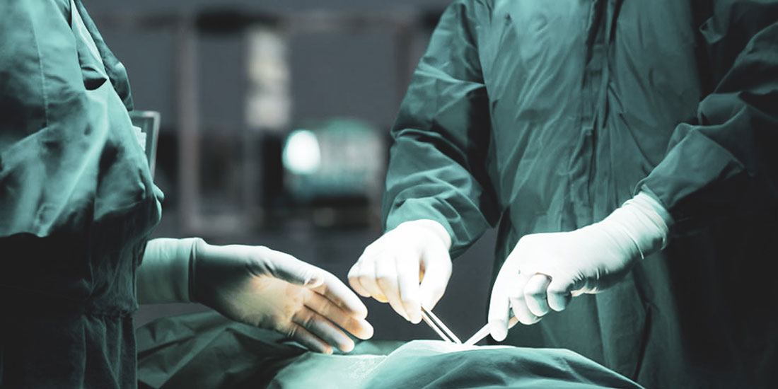
Surgical pathology of the liver poses intricate and multifaceted hurdles for surgical pathologists. You work with different specimens and many questions arise as you plan how to perform a successful analysis. Generally, you’ll need to use a needle-core biopsy or a larger wedge biopsy; a partial hepatectomy might also be required. The outcome and considerations vary greatly based on the unique characteristics of individual cases. The primary doctor who treats the patient needs an accurate diagnosis with all the details of the patient’s medical history. That’s why the collaboration of surgical pathologists with the whole medical team is crucial. It is imperative to establish the necessary conditions to ensure the attainment of the most precise and accurate diagnosis irrespective of the type of liver specimen under examination. That means satisfying all the technical requirements, primarily having the adequate size of the specimen and the best technical preparation. The following text will explore the importance of liver biopsy in the context of surgical pathology and the prerequisites for its successful performance.
How to Perform Liver Biopsy?
There are generally two types of liver biopsies: needle-core biopsy and wedge biopsy, which is larger in size. No matter what form of biopsy is to be conducted, the specimen should be measured and submitted in its entirety for routine histology. It’s worth mentioning that thin-core biopsies are particularly susceptible to drying out. Unless special examinations are needed, the biopsies should be placed in fixative in the operating room. The core biopsy can be embedded whole while the wedge biopsy may be thick enough to warrant sectioning before submitting it to the histology laboratory. When sectioning the wedge biopsy, you should identify the smooth capsule, and then cut the liver at 0.2-cm intervals perpendicular to its surface. When it comes to histologic evaluation, multiple slides should be prepared from each tissue block for histologic evaluation. Surgical pathologists usually prefer step sections to serial sections because intervening sections need to be available for special stains. If a storage disease is suspected, then a small portion of the biopsy should be placed in glutaraldehyde for electron microscopy.
Stay Ahead with Insights from Precipoint!
Welcome to our newsletter! Be the first to know about our latest products, services, webinars, and happenings in PreciPoint. Don't miss out on this opportunity to stay informed. Subscribe to our newsletter today!
By clicking “Subscribe”, you agree to our privacy policy.
When to Use Partial Hepatectomy?
Sometimes in surgical pathology, focal lesions of the liver need to be removed by partial liver resections. This procedure is called partial hepatectomy. The extent of these resections can vary from small wedges to the removal of the entire lobe. Regardless of its size, the partial liver resection is not structurally complex. It consists typically of a focal lesion surrounded by a variable rim of non-neoplastic liver parenchyma. Several faces of the specimen are covered by a peritoneal lining, but at least one surface shows exposed hepatic parenchyma. This exposed surface is the surgical resection margin that must be correctly identified and oriented. Sometimes, the identification of the resection margin can be problematic due to the smooth contour of the peritoneal-lined liver capsule. The resection plane exposes liver parenchyma which may be fragmented or bloody and show a cautery effect. However, because of the paucity of anatomic landmarks in these limited resections, the orientation of the margin can be very difficult. For example, if the surgeon needs to know the precise location at which the tumor involves or approaches the surgical margin, orientation will require the surgeon’s assistance.
Once the margin is identified and the specimen oriented, you need to weigh the specimen and measure it in each dimension. It is also important to examine its contours and surfaces. A bulge in the surface of the liver or retraction of the serosa can help localize an intraparenchymal mass. Livers resected for traumatic ruptures should be carefully examined for lacerations of the capsule. Then you should rinse the blood from the cut margin, blot it with paper towels, and then ink the dry resection margin. The initial section should pass through the center of the tumor to demonstrate the close approach of the tumor to the resection margin. You should then continue the section of the liver parallel to this first cut at thin intervals. It is also necessary to record the number, size, location, color, consistency, and circumscription of all lesions. You should also note the presence or absence of necrosis, hemorrhage, and scarring. It is necessary to measure the distance from all lesions to the surgical resection margin.
Evaluation of Liver Biopsies
Liver biopsies taken for non-neoplastic diseases are relatively uncommon in general pathology laboratories and are often received with no, incomplete, or occasionally misleading histories and minimal laboratory data. You need to generate a reasonable differential diagnosis and become comfortable communicating with the gastroenterologist or hepatologist about the result of the biopsy.
Successful Analysis of Liver Biopsy
The key to successfully interpreting a liver biopsy lies in obtaining an adequate specimen size and employing optimal technical preparation methods. Adequacy of size largely depends on the process being evaluated. If one is looking at metastatic carcinoma and there is a tumor present, the size of the tumor is crucial. However, more tissues are needed to rule out some diseases such as primary biliary cirrhosis. For the most chronic diseases, a minimum of 1.5 cm is necessary, although, some diseases such as hepatitis C suggest significant staging error with less than 2.5 cm.
The application of specific stains depends upon the circumstances and individual preferences of the surgical pathologists, considering their unique perspectives and expertise. Usually, the routine consists of hematoxylin and eosin, trichrome and iron preparations. Iron stain is used to determine hemochromatosis. If the biopsy is assessed carefully on H&E staining, significant iron deposition as the granular golden-brown pigment is visible without the iron stain, rendering the stain optional if one diligently searches for pigment. Remember that small amounts of iron may be difficult to identify, and lipofuscin pigment is common in biopsies and simulates iron on H&E stain. Generally, it is centrilobular rather than periportal, as iron is in hemochromatosis. The best approach to the interpretation of the biopsy is to review the slide without any knowledge of clinical or laboratory data. A systematic evaluation of portal tracts, lobules, and central veins should be undertaken, assessing features such as degree and type of inflammation, necrosis, and fibrosis. After noting the findings, a diagnosis should be determined.
Optical Imaging for Rapid Evaluation of Tissues
Optical imaging techniques are currently available for imaging tissues without the need for any type of extensive tissue preparation. Confocal fluorescence microscopy is utilized for ex vivo examination of tissues obtained from surgical resections of the breast, lung, kidney, or liver. In the process, you acquire CFM images very quickly and easily. These images resemble hematoxylin-eosin sections, and that suggests that the CFM platform has exceptional potential for use in surgical pathology practice. Optical imaging encompasses several techniques that can be used for non-invasive imaging of biological tissue specimens. This method uses light in the visible spectrum and harnesses the special properties of photons to obtain detailed images of tissue without exposure to harmful radiation. If you need to perform repeated procedures over time, optical imaging platforms are safer, faster, and well-adapted. You can also perform the targeted biopsy more easily and effectively with digital microscopes and scanners such as M8 and O8 and perform better categorization of mucosal changes.
Conclusion
Surgical pathology of the liver encompasses several methods and techniques to come up with an accurate diagnosis. Among the approaches, a biopsy stands out as a significant procedure that entails a variety of techniques. The methods are tailored to the unique circumstances of each case, encompassing the required specimen size ranging from small wedges to the entire lobe. Successful liver biopsy also involves the identification of the margin and specimen, and the measure of the same specimen. The most important thing in the whole biopsy of the liver specimen is the preparation of the specimen for analysis. The next critical task is the technical preparation for the analysis. If you are looking at metastatic carcinoma and there is a tumor present, the size of the tumor is crucial. After assessing all the features of the specimen, you write a report and help in setting up the diagnosis. Advancements bring a lot of advantages to surgical pathologists. Optical imaging is one such technique that has emerged as a valuable tool. The results obtained by employing cutting-edge devices like PreciPoint’s M8 and O8 digital microscopes and scanners can reach remarkable levels of quality and precision.