

Breast cancer is by far the most common cancer in women: according to estimates by the Robert Koch Institute, 69,700 women were recently affected by it in Germany, and more than 18,000 women died of it. One of the most common treatment approaches is a combination of radiotherapy and surgery. If surgery is possible, the surgeon may remove the tumor and send it to the pathologist’s laboratory. However, this step is often complicated due to a lack of pathology staff.
For this purpose, the company PreciPoint has developed a fully motorized brightfield microscope that allows remote access to microscopic samples in seconds – making it easier for pathologists to obtain a second opinion and work collaboratively.
Therapeutic options for breast cancer
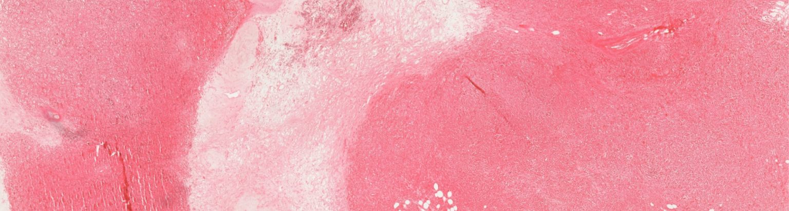
Currently, one in eight women will develop breast cancer in their lifetime. Therapeutic options range from radiation and hormone therapy to chemotherapy. In most cases is surgery indispensable but can now be performed in a breast-conserving way. It is important to ensure that the margins of the removed tumor are free of cancer cells so that the cancer does not spread again. To ensure this, the breast cancer guideline program recommends the intraoperative frozen section: part of the removed tissue is brought to the laboratory during the operation. It is shock-frozen and cut in very thin sections.
A pathologist assesses these sections under the microscope and informs the surgeon whether the edges of the tumor are free of cancer or not. Due to the shortage of pathologists worldwide, the frozen section is often omitted, and the removed tissue is only examined microscopically after the operation. As a result, some operations have to be repeated, which puts physical strain on the patients and causes high costs.
Our contribution
The company PreciPoint has developed a solution for this that makes it possible to obtain a second opinion easily and quickly and to facilitate cooperation between pathologists and surgeons. The system, which is currently being developed and certified, is a robotic brightfield microscope that produces a live image of microscopic specimens in seconds. The image is transmitted to a screen, and the system is operated without delay via a touch screen or by mouse click.
Another advantage over a conventional microscope is that the PreciPoint solution captures an overall image of the sample, unlike a conventional microscope where only a single, small section can be viewed through the eyepiece at high magnification. Once PreciPoint has successfully completed the development and certification processes, the company plans to deploy the solution in laboratories and clinics around the world.


