
Synopsis
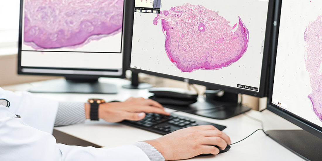
Image acquisition is critical for virtual microscopy to take the quality of digital laboratory work to the next level. As virtual microscopy becomes a daily routine for digital pathologists, there is a need to standardize image acquisition and display. The standards should meet the daily needs of the digital laboratory, including optimization of results and high-quality output. You need to effectively adjust the brightness, contrast, and magnification, and use appropriate camera settings to ensure maximum diagnostic integrity. Let’s explore the key elements that make up optimal digital image acquisition.
What Does Image Acquisition Mean in Virtual Microscopy?
Image acquisition is one of the necessities of virtual microscopy. The development of next-generation slide scanners has made virtual microscopy a routine part of everyday digital pathology lab workflow. Therefore, image acquisition and display requirements must be met in a standardized format to ensure consistency throughout the process. Virtual chips require the processing of large amounts of data, so image compression is also an important consideration.
The Right Combination
The maximum available magnification, diagnostic security, scanning time, and data compression are factors that affect results depending on the intensity of their use. Typically, slides can be digitized at different quality levels such as 100%, 90%, 85%, 80%, 70%, 60%, and lower. The quality level of slides impacts setting up the right diagnostic requirements.
Achieve High-quality Image
Some researchers believe that high-quality images can only be obtained at a quality rate of 40% or higher. The higher the magnification, the better the results. In addition, maintaining a high-quality level of approximately 90% while reducing the amount of data reduces the time required for digitization. In addition, the lower the amount of data, the lower the time required. The time required for digitization also depends on the speed of the network.
Balancing the Ratio
The compression rate is about 20% for a 90% quality step and drops further to 8% for a 10% quality step. Thus, a pixel resolution of 0.2 x 0.2 square micrometers and a quality step of 40 to 80% is acceptable for a high-quality image. A resolution of more than 4 megapixels and a contrast ratio of more than 3,000 are ongoing technical requirements for virtual slideshows. Since .jpg compression is a flexible compression standard, all slide scanners must use the standard compression mode.
Best Image Acquisition
Virtual microscopy standards ensure a safe and collaborative environment for pathologists. In virtual microscopy, digital image acquisition automates the entire digitization process. Digital monochrome and color cameras allow for transmitted light, providing you with high-quality results. In fluorescence microscopy, monochrome cameras are often the preferred acquisition method.
Technical Requirements
Virtual microscopes can digitize large digital images at high resolution for both brightfield and fluorescence. The quality of a digitized image can be measured by three metrics: sharpness, contrast, and focus. In general, optimal hardware setup and software acquisition are key to obtaining the best images. In brain dissections, prostate biopsies and lung biopsies, magnification from 2.5x to 63x produces the best image quality.
Digital Image Acquisition and Artificial Intelligence
Successful image acquisition also involves the basic fact of developing artificial intelligence (AI) models for deep learning. If you work in the AI-integrated virtual microscopy field, you may have encountered situations where you have made extensive annotations for a learning model, but the results are confusing, and you have to repeat all the annotations.
Conditions Apply
While many pathologists have extensive practical experience in providing scientifically accurate annotations from a pathology perspective, they may not always meet computer vision standards. As a result, these annotations may be less accurate or valid for computer vision analysis, resulting in a loss of accuracy or reliability. To ensure optimal capture quality and minimize problems, it is necessary to follow certain rules regarding clean classes, annotation size, error handling, and maintaining consistency.
- Clean classes: Classes must be clean and tidy to avoid pixel confusion in the annotations. For example, if the annotations are too small, it will not be possible to show the heterogeneity of the tissue sample or the transition of one appearance of the tissue into the other.
- Annotation size: The size of the annotations partially depends on the heterogeneity of the tissue under study. When developing deep learning models, it is important to consider the potential challenges posed by the limited presence of pathological structures in medical data. Inherent imbalances in datasets must be addressed and corrected accordingly.
- Class imbalance: Classification imbalance occurs when underrepresented categories are overrepresented or overrepresented categories are underrepresented. Correcting this requires adjusting all information provided by direct observation and measurement during the modeling process.
- Consistency: You need to go back and align everything with the project requirements After the project and all the necessary interpretation cycles have been completed. It is important to be alert to biases and offsets that occur during this process, so model training must be revisited and refined before finalizing the model.
Conclusion
Optimizing digital image acquisition in virtual microscopy means an accurate, efficient, and precise workflow and safer diagnostics. To build the right artificial intelligence (AI) learning models and develop technology, standardization is necessary for optimizing image acquisition. Attention needs to be paid to basic requirements such as magnification, brightness, contrast, and image compression to implement standardization. Image quality can also be improved to many folds by using the right software and AI learning models. It is necessary to carefully consider factors such as clean classes, annotation size, class imbalance, and consistency. Image acquisition for virtual microscopy must be optimized to improve efficiency and quality. This is made easier through the use of best-in-class software and hardware solutions, and PreciPoint specializes in providing microscopes, digital scanners, services, and solutions that precisely meet your specific requirements, allowing you to get the highest performance from your work.