
Synopsis
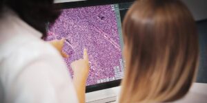
Nucleus segmentation is one of the most important activities in digital pathology. It involves identifying and defining the boundaries of the nucleus. Nuclear fragmentation facilitates cancer diagnosis, progression, and prognosis. Computer-based methods take modern digital pathology to the next level, making diagnosis and analysis more efficient and quality-oriented. Nucleus segmentation methods overcome challenges such as significant changes in nuclear appearance due to staining, tissue preparation, and imaging environment. Modern techniques help address inconsistencies and control disturbances such as noise, debris, and folds in the specimen. This article explains how nucleus segmentation is evolving in modern digital pathology workflow.
Why Do We Need Nucleus Segmentation?
Nuclei segmentation represents a crucial step in many applications and practices, such as cancer detection, grading, and prognosis. Segmenting cell nuclei in histopathology images is the preliminary step in analyzing current imaging data for biological and biomedical purposes. The fundamental property of nucleus segmentation is to analyze, diagnose, and grade cancerous cells. This is done through cell counting, movement tracking, computational pathology, cytometric analysis, computer-aided diagnosis, and morphological study. During nuclei segmentation, you identify and delineate the boundaries of the nuclei.
Meticulous Segmentation
Accurate nuclei segmentation is important because the morphology and location of nuclei are critical for tissue characterization and diagnostic quantification. Cancer diagnosis uses the size, shape, and distribution of nuclei as indicators of malignancy. The diagnosis, classification, and presentation of cancer are largely dependent on the quality or precision of nuclear segmentation. Accurate segmentation of nuclei helps to identify cancer cells faster and distinguish between malignant and benign cells.
Technical Development
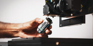
In recent years, computational techniques have developed rapidly in digital pathology, and cancer detection, segmentation, and classification. Technological advances play an important role in reducing human intervention, incorporating relevant second opinions, and searchable medical information.
Challenges Faced During Nucleus Segmentation
One of the major challenges in nucleus segmentation is the high variability of nuclei due to differences in staining, tissue preparation, and imaging conditions. Additionally, nuclei can overlap with each other, making it difficult to separate them. Another challenge is the presence of artifacts such as noise, debris, and folds in the tissue. These disturbances can interfere with accurate segmentation.
Nucleus Segmentation Techniques
Several techniques address the challenges arising during nucleus segmentation. These techniques include different methods to handle variations, image regions, and nuclei segments. Each of these methods offers unique strengths and can be tailored to specific applications and datasets. The methods are:
Thresholding
That is a simple method that involves setting up a threshold value and classifying pixels as nuclei or backgrounds based on their intensity values. It is important to note that this method is sensitive to variations regarding staining and illumination and can lead to over- or under-segmentation.
Watershed Segmentation
This is a region-based method that segments an image into regions based on the gradient values of the image. The boundaries of the regions are determined by the watershed lines, and these lines separate the catchment basins of the gradient peaks. Watershed segmentation is robust to staining and illumination, allowing for accurate segmentation of nuclei. However, this method also poses a challenge as it is sensitive to noise and can lead to over-segmentation.
Machine learning-based methods
These techniques were developed to overcome the challenges associated with thresholding or watershed segmentation. Machine learning-based methods involve supervised learning algorithms to train a model to classify nuclei in pathological images. The model is trained on a dataset of annotated images and learns to identify features necessary for cellular segmentation. However, obtaining a database can be time-consuming and expensive. Once the model is trained, it can be used to classify nuclei in new images. These techniques can be used to observe changes in the appearance and artifacts of images.
| Technique | Advantages | Limitations | |
|---|---|---|---|
|
Thresholding |
Sets up a threshold value and classifies pixels as nuclei or backgrounds based on their intensity values. |
|
|
|
Watershed Segmentation |
Segments an image into regions based on the gradient magnitude of the image. |
|
|
|
Machine Learning-based Methods |
Supervised learning algorithms train a model to segment nuclei. |
|
|
Nucleus Segmentation Applications
Nucleus segmentation can help study the structure of neurons. Changes in the morphology of the nuclei may indicate abnormalities. When you do the accurate segmentation of the nuclei, you can facilitate the quantification of various morphological features such as size, shape, texture, and distribution. You can utilize these features to study tissue morphology and the effects of disease on tissue specimens you observe and analyze.
Conclusion
Nucleus segmentation is an important aspect of digital pathology. By applying nucleus segmentation, you can research, analyze, and view samples, even when working on the most complex cases. Thresholding, watershed segmentation, and machine learning-based methods provide details that might not be possible to achieve with traditional techniques. The modern-day techniques help you make decisions related to your research, diagnosis, and finally, the further treatment of your patient. Most importantly, image analysis is crucial in nucleus segmentation. When you take digital microscopes and AI simultaneously, using AI-powered image analysis algorithms, you create an environment that benefits from increased accuracy and precision due to advanced materials, as well as greater flexibility.