
Non-neoplastic Gastrointestinal Diseases: Surgical Pathology of Gastritis
Synopsis Establishing a diagnosis of gastritis within non-neoplastic gastrointestinal conditions can pose significant challenges. Gastritis, the disease characterized by the
All the news, insider reports, commentary and excitement from the world of Digital Microscopy from PreciPoint.
news and interesting facts about microscopy and research. Industry events, trade shows and events from the world of microscopy!

Synopsis Establishing a diagnosis of gastritis within non-neoplastic gastrointestinal conditions can pose significant challenges. Gastritis, the disease characterized by the

Synopsis Nucleus segmentation is one of the most important activities in digital pathology. It involves identifying and defining the boundaries
Synopsis Traditional surgical pathology workflow was dependent on manual processes and often it faced challenges arising from human errors and
Synopsis Magnification and resolution are the two sides of the same coin in digital pathology. As a pathologist, you might
Synopsis The microscopic world offers a variety of opportunities for surgical pathologists. By mastering the basic techniques of digital microscopy,

Synopsis Quality control is one of the essential parts of digital pathology and histopathology. Especially in histopathology, not many medical
Overcoming digitization challenges in pathology involves addressing equipment costs, standardizing protocols, integrating complex systems, and ensuring technology stability.

Telepathology in digital pathology enables remote consultation for rapid diagnosis and collaboration, improving patient care in underserved areas.

Digital microscopes for higher education should have high magnification, excellent image resolution, built-in cameras, proper lighting, durability, and user-friendly software.
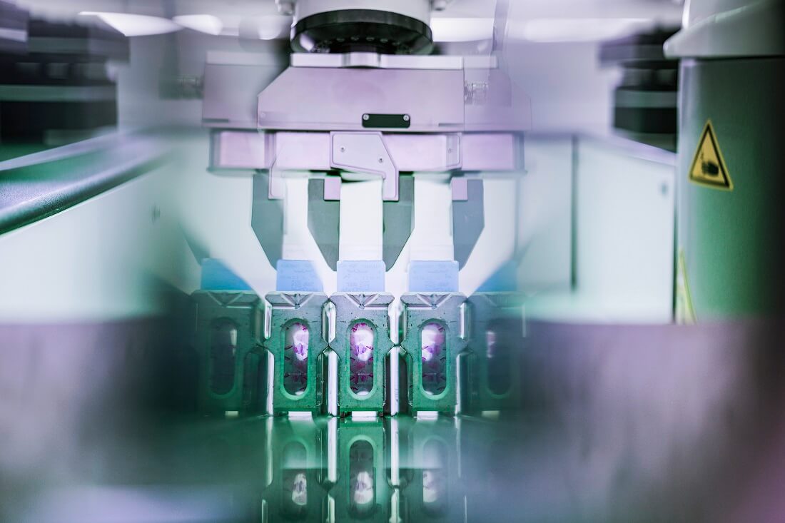
Implementing digital pathology involves a systematic approach, including team integration, resource optimization, workflow validation, staff training, system comparison, cost evaluation,
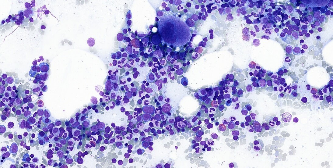
Paperless quality control for bone marrow testing enhances accuracy and efficiency through digital data entry, image processing, and reporting.
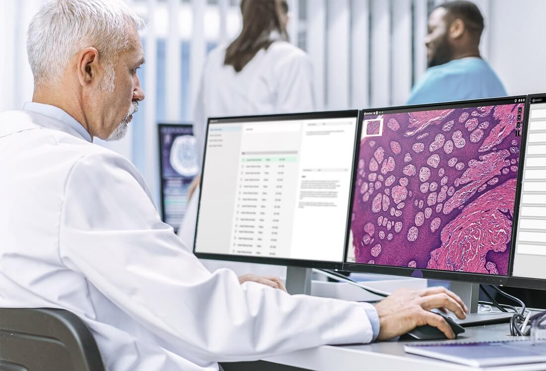
The reliability of digital microscopes is crucial for accurate diagnostics, necessitating regular maintenance, proper lighting, and up-to-date software.
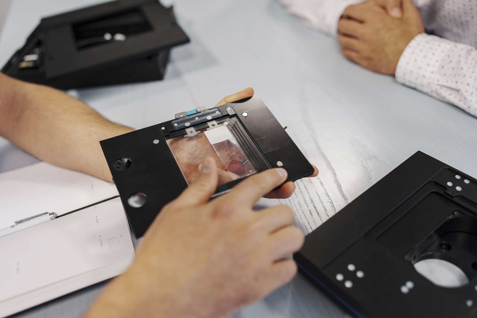
Digital microscopes increase intraoperative consultations by enabling real-time remote viewing and collaboration, improving decision-making during surgeries.

Digital pathology in intraoperative procedures faces challenges like integration, scanning speed, image quality, file management, and regulatory approvals.

Digitizing pathology workflows faces challenges like image quality, data management, system integration, workflow compatibility, and regulatory compliance.

Digital microscopes enhance pathology with rapid, high-quality imaging, improving diagnosis speed and accuracy, and enabling remote consultations.

During telepathology practice, the shortage of medical experts is one of the greatest challenges that developing countries are facing.

Choosing an appropriate research microscope is fundamental for high-quality digital microscopy.

The microscope’s resolution greatly influences the value and effectiveness of the pathological process.

Telepathology through the lens of virtual microscopy doesn’t just hold potential; it’s a blazing trail of progress.

Machine learning (ML) algorithms integrated with whole slide imaging have firmly established their presence.

Misconceptions surrounding digital pathology continue to impede its widespread adoption globally.

The digitalization of continuing medical education (CME) marks a transformative era fostering personalized, engaging learning experiences.

In the dynamic space of microscopy, a transformative paradigm shift is underway, fueled by the rapid integration.
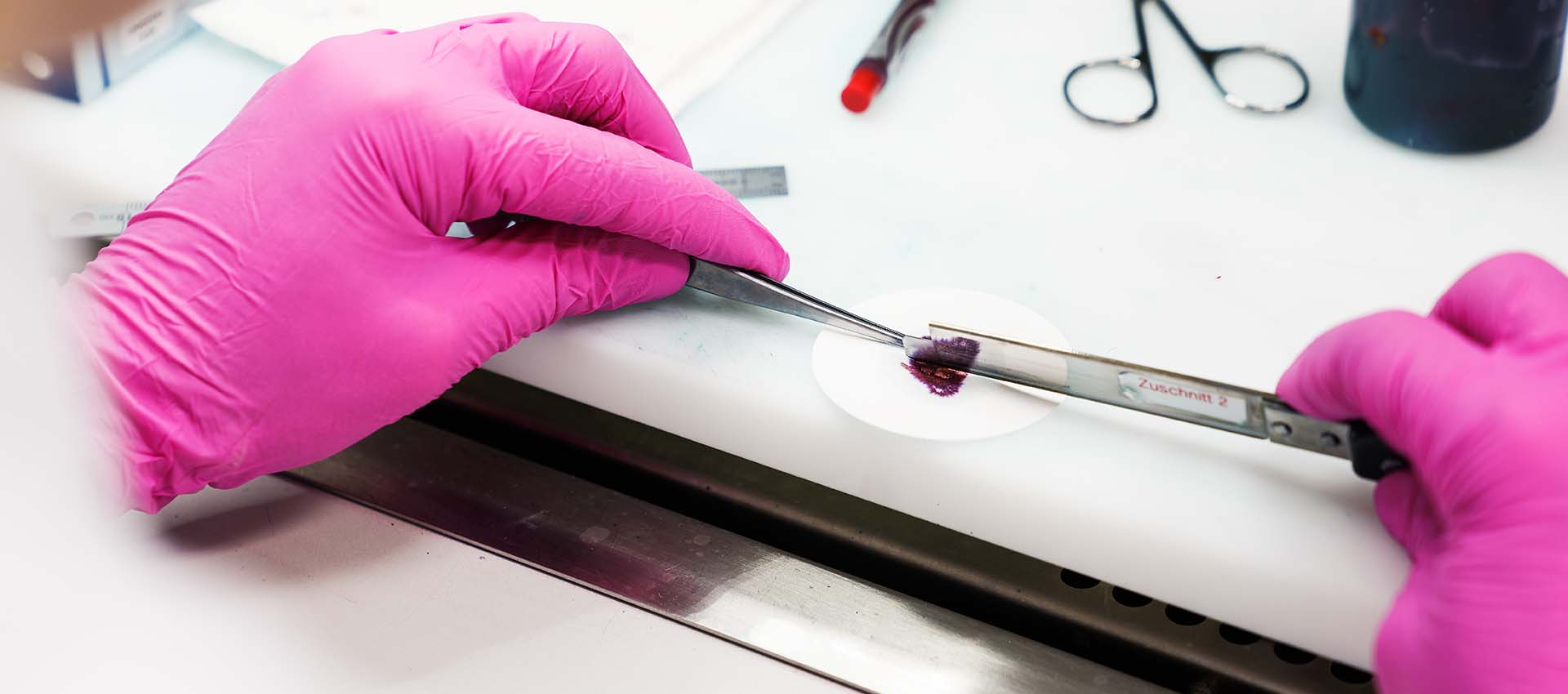
In pathology, traditional methods, such as research by using a conventional microscope, aren’t the most used techniques anymore.
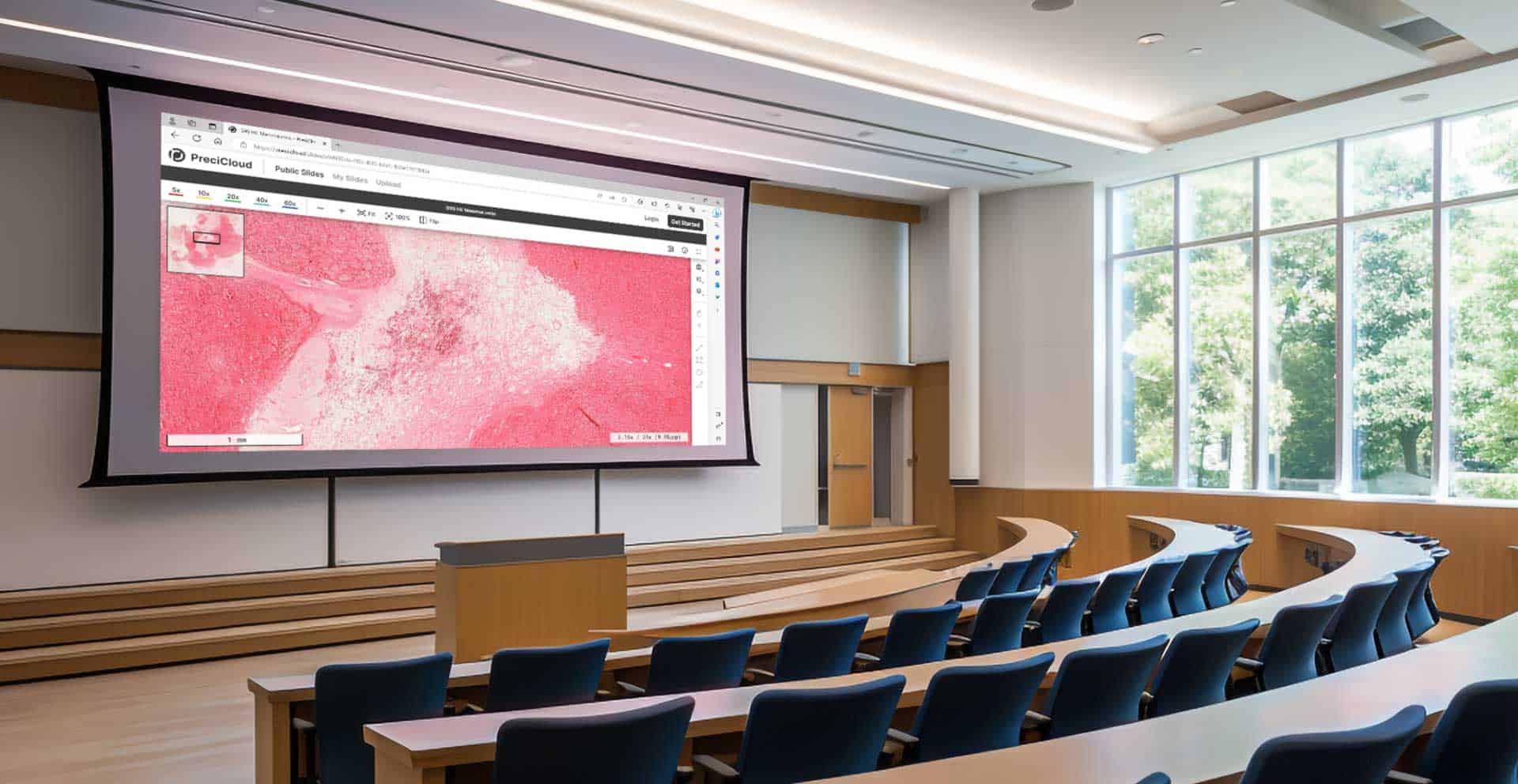
Discover how virtual microscopy revolutionizes pathology education by enabling interactive, accessible learning from anywhere.

Explore how deep learning enhances breast cancer diagnosis through digital pathology advancements.

September is Prostate Cancer Awareness Month, the right time to delve into its fundamentals.

Brightfield microscopy is one of the oldest and most widely used techniques among researchers and medical experts.
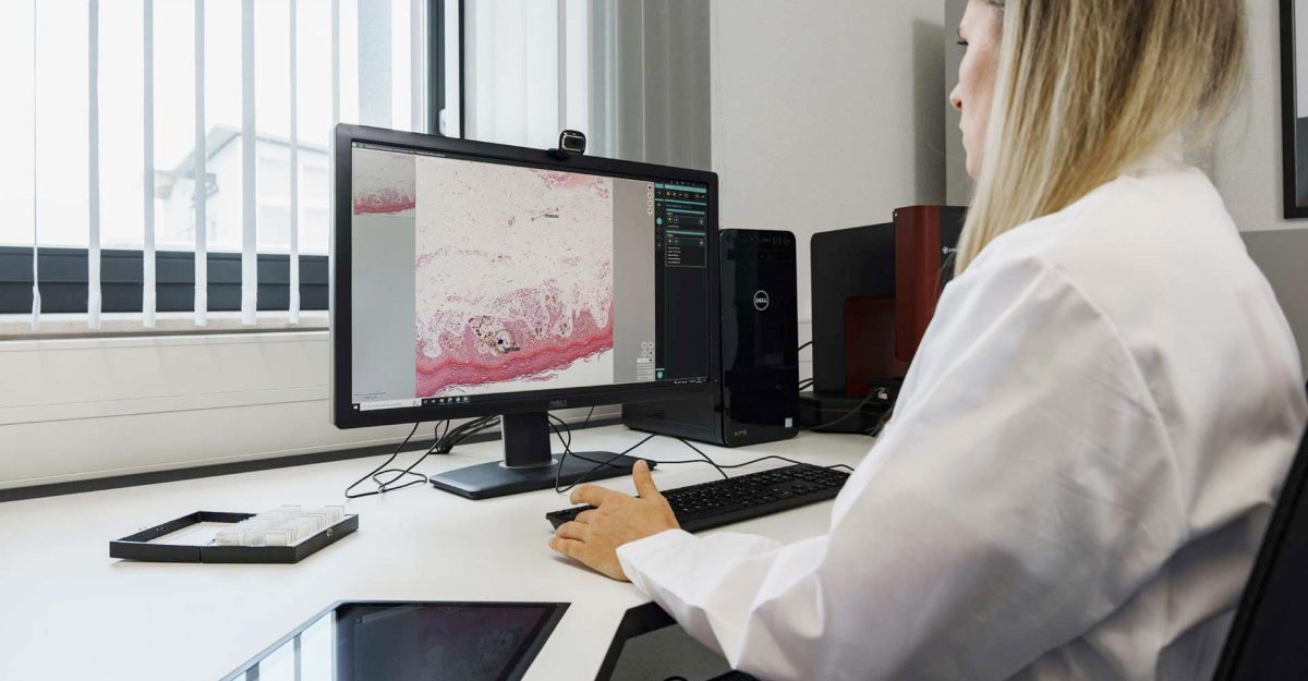
Expert tips to enhance digital microscope use include proper resolution, lighting, and regular maintenance for optimal results.
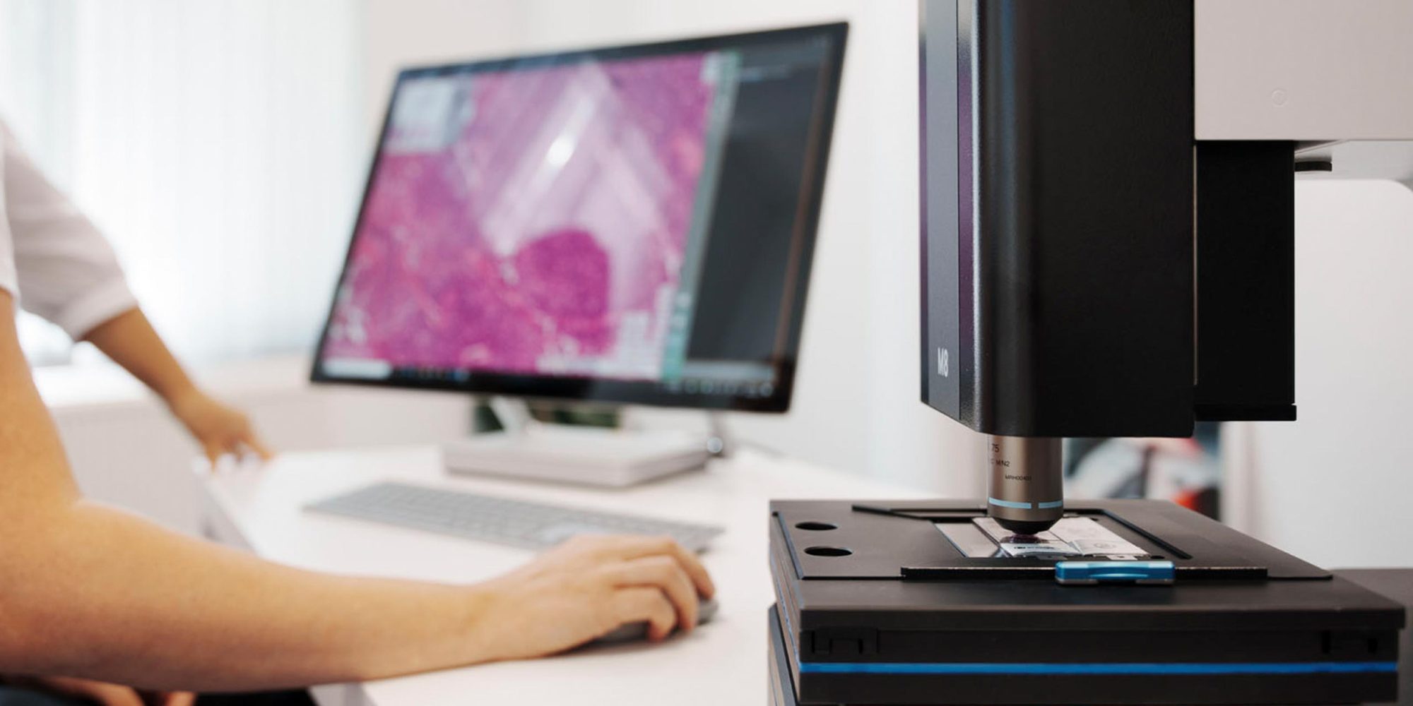
Digital microscopes enhance analysis and collaboration by simplifying data sharing and reducing physical strain during extended use.

Digital microscopy is transforming pathology education by standardizing materials and increasing image accessibility.
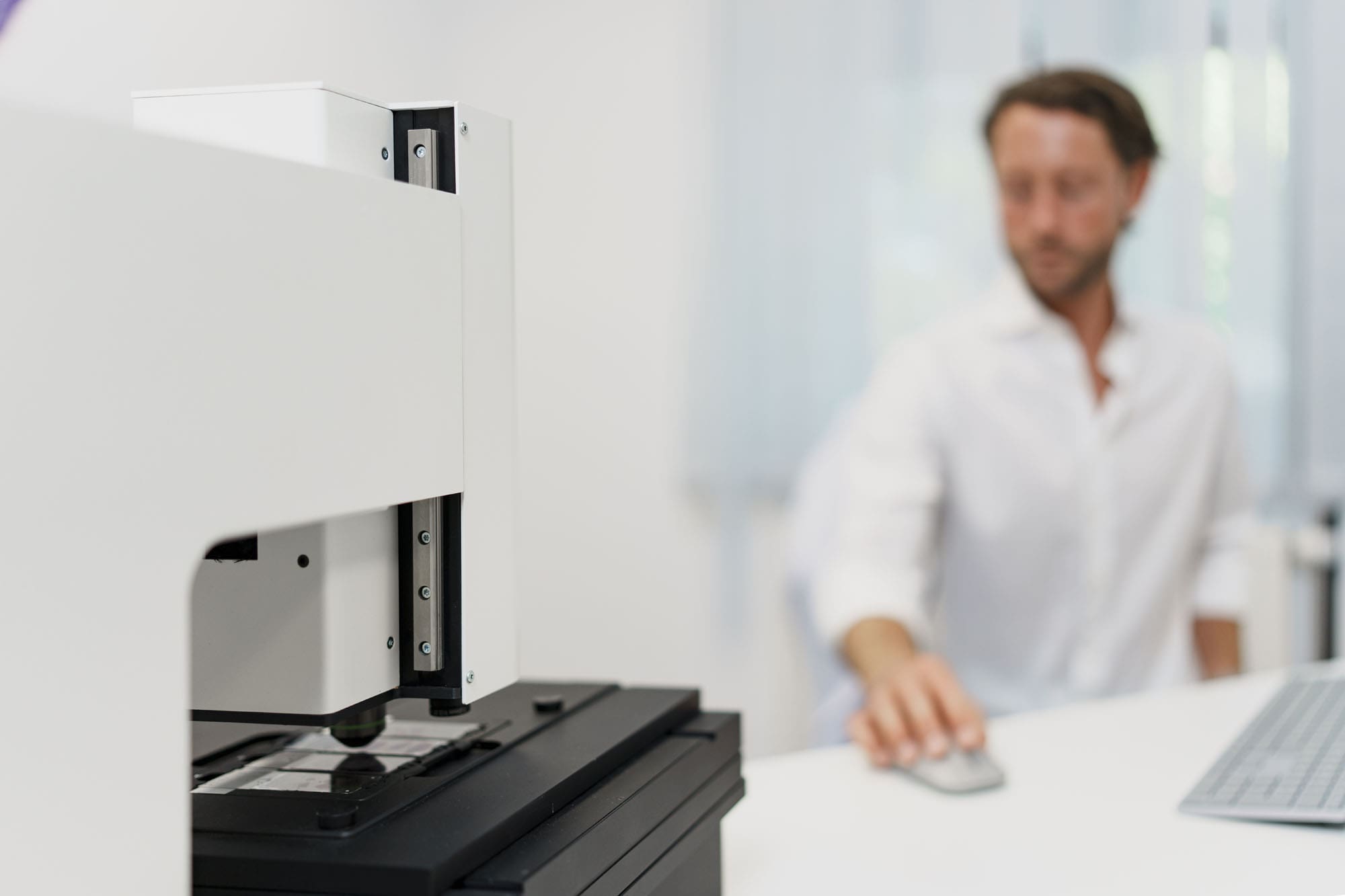
Digital pathology streamlines cancer diagnosis with AI-driven tools for faster, more accurate assessments.

Digital microscopes enhance cancer diagnosis accuracy through high-resolution image analysis and AI integration.

Intraoperative frozen section examinations reduce reoperations in breast cancer by providing quicker diagnostic results.
Digital frozen section analysis faces challenges in speed and quality due to complex slide characteristics.

Digital microscopy facilitates real-time surgical decisions by connecting surgeons and pathologists.

Digitizing intraoperative consultations can accelerate second opinions, save pathologists’ time, and expand global access to pathology services.
Optimal slide preparation for digital microscopy requires careful placement and absence of folds or air bubbles.
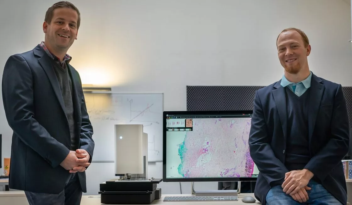
A Bavarian startup’s innovative microscope aims to streamline breast cancer surgeries by eliminating the need for physical slide transport.
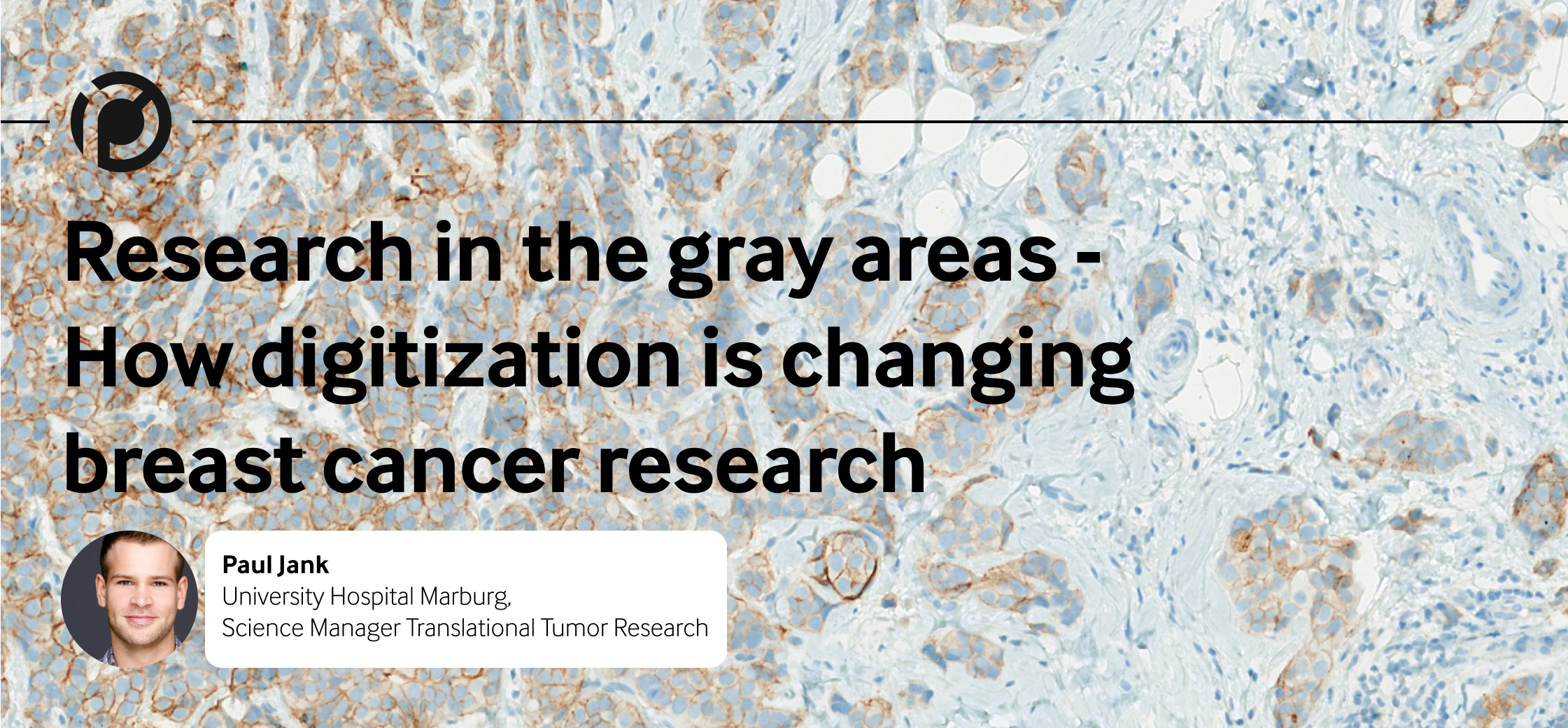
Digitization is reshaping breast cancer research by supporting the development of personalized treatment approaches.

Digitization in brewing is highlighted as a significant advancement, enhancing quality control and operational efficiency.

Digitization in research dramatically speeds up access and analysis of samples, enhancing collaboration and AI integration for biomarker studies.
PreciPoint and Nikon Europe have partnered to distribute digital microscopes across multiple European countries, enhancing lab digitization.

Digital microscopy improves lecture quality by enabling simultaneous access to digital slides, facilitating more interactive and collaborative learning environments.

AI-enhanced digital microscopy selectively scans only relevant tissue, improving image quality and reducing data storage needs.
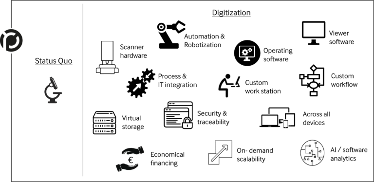
Digital laboratories boost efficiency by integrating various digital technologies and devices, which not only improves workflow but also increases quality
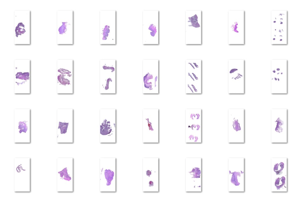
The PathoScan project aims to fully digitize pathology labs with a focus on automated and scalable sample processing, increasing throughput


Welcome to our newsletter! Be the first to know about our latest products, services, webinars, and happenings in PreciPoint. Don’t miss out on this opportunity to stay informed. Subscribe to our newsletter today!
By clicking “Subscribe”, you agree to our privacy policy.