
How to save a microscopy overview image file?
- To generate thumbnail images of slides
- In general, for documentation e.g.: marking of scan areas
- Comparison with other image-based analyzes for assigning signals from specific regions to the tissue context e.g., HE-stained tissue compared to chromogenic in situ hybridization (CISH) or immunohistochemistry (IHC)
- Saving selection/marking(s) of areas within the overview image for further analysis, e.g.
mass spectrometry - AI training of overview images to e.g., automatically define useful scan areas (reducing of scanning times)
Stay Ahead with Insights from Precipoint!
Welcome to our newsletter! Be the first to know about our latest products, services, webinars, and happenings in PreciPoint. Don't miss out on this opportunity to stay informed. Subscribe to our newsletter today!
By clicking “Subscribe”, you agree to our privacy policy.
How to save a microscopy slide overview image?
As an existing customer, please write us your wish to use this feature via email or portal. We then activate this feature via remote session. Upon request, new customers can get the feature directly with the delivered software.
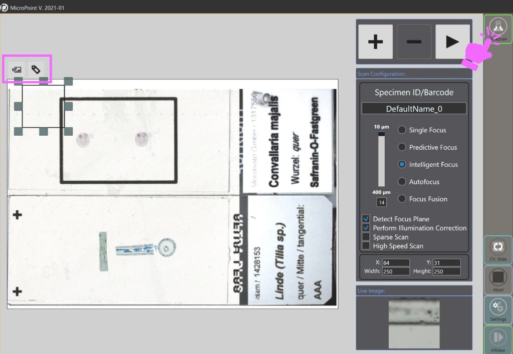
If the feature is activated, the following buttons can be selected under the Slide Scan mode

To save the overview image, click on the left button (to save the label, use the right button)
The overview image can be saved in the following formats: JPEC, Bitmap, PNG or TIFF.
Quick Note: This feature will soon be directly enabled in the Slide can mode and will no longer need to be activated separately.
This feature is available in the MicroPoint Software for the following devices:
- M8
- O8
- Fritz