
Synopsis
The microscopic world offers a variety of opportunities for surgical pathologists. By mastering the basic techniques of digital microscopy, your laboratory can embark on the journey toward unparalleled levels of quality and excellence. As a user of digital microscopes, you need to master basic concepts namely resolution, image size, DPI and PPI. These elements are essential to unlocking the full potential of digital microscopy and achieving your and your patients’ satisfaction. In this article, we discuss the main aspects of digital microscopy and emphasize the importance of resolution, image size, DPI and PPI. By examining these basic elements, we aim to provide a comprehensive understanding of their importance, while addressing other important aspects of the field.
Unraveling the Potential of Digital Microscopy
In recent years, digital microscopy has transformed the world of surgical pathology. For pathologists, the microscopic world is a fascinating canvas and an opportunity to unlock the mysteries of life on a small scale. Digital microscopy provides powerful tools for capturing, analyzing, and sharing high-resolution microscopic images. However, to fully utilize the potential of a digital microscope, one must understand basic concepts such as resolution, image size, DPI (dots per inch), and PPI (pixels per inch).
Mastering the Image Resolution
Resolution is the backbone of any digital system. Microscopy allows you to distinguish fine details and separate closely spaced objects in an image. You primarily work with the numerical aperture (NA) of the microscope’s objective lens and the wavelength of the light used for illumination. By using larger numerical apertures and shorter wavelengths, higher resolution images can be achieved.
Image Resolution in Surgical Pathology
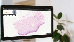
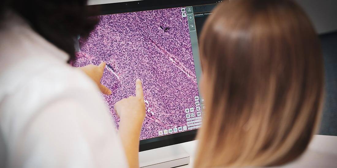
In surgical pathology, resolution is critical for capturing finer details and improving image quality. With the PreciPoint M8 you can easily and effortlessly zoom in on the digital slide. The intuitive operating concept enables consistent navigation. The software offers functions such as annotations, measurements, multiple fields of view and much more that provide valuable support to your work.
Achieve High Magnification
High magnification is the easiest to achieve. All you must do is take a photo of the probe and zoom in. While the resolution of a microscope is primarily determined by the optical system, various techniques and considerations can help maximize resolution. Enhancing resolution in the framework of surgical pathology is crucial when capturing fine detail:
- Choose the Right Microscope: If you select a microscope with a high NA, you can achieve better resolution. A higher NA allows you to capture the light rays from steeper angles, resulting in improved image quality and increased resolution.
- Optimize Illumination: Proper illumination is the key to enhancing resolution. You should make sure that your microscope is equipped with a bright, uniform light source. Technologies like Köhler illumination can help improve lighting by aligning the light path and reducing aberrations that can affect accuracy.
- Reduce Aberrations: Aberrations, such as spherical aberration or chromatic aberration, can negatively impact the resolution. Therefore, you need to check and clean the optics of your microscope regularly to maintain its performance. Using high-quality lenses and implementing adaptive optics, whether through software or hardware solutions, improves accuracy by correcting aberrations.
- Use Immersion Techniques: Immersion techniques, such as oil immersion or water immersion, can significantly improve resolution. By reducing the refractive index mismatch between the sample and the objective lens, immersion techniques reduce light scattering and refraction, resulting in better resolution. O8 is the right solution for the application of immersion techniques. O8 is the ideal device for researchers. With up to 100x air and oil lenses, you can scan entire slides and specific areas of interest.
- Image Stacking and Deconvolution: Image stacking involves taking a series of images at different focal planes and then combining them into one clear image. This technique improves the depth of field and overall resolution by compensating for the limited depth of focus in microscopy. Additionally, you can also apply deconvolution algorithms to remove out-of-focus light and further improve resolution.
- Post-processing Techniques: You need to use image processing software to improve resolution after taking photos. Techniques such as sharpening, noise removal, and noise reduction can help improve the clarity and detail of the image. These techniques are important because they allow you to avoid introducing artifacts or losing valuable information.
- Consider Super-resolution Techniques: Some techniques go beyond light diffraction and allow the visualization of very fine details. These techniques include structural illumination microscopy (SIM), stochastic optical reconstruction microscopy (STORM), and stochastic emission depletion microscopy (STED). They can be used to carry out high-resolution digital microscope experiments.
- Optimal Camera Settings: You need to make sure that your camera settings are optimized to capture high-resolution photos. You need to adjust the exposure time and other relevant parameters to achieve the best signal-to-noise ratio. You can also use the highest depth bit available to capture a larger grayscale range and retain more detail in the image.
Importance of Image Managing
Resolution is closely related to the concept of image size. Image size includes the dimensions of the image file that represents the captured image. Typically expressed in pixels, they are the individual building blocks of a digital image. With a digital microscope, it is necessary to balance image size and resolution. Higher resolution requires more pixels to capture fine details, resulting in larger file sizes. Conversely, a lower pixel count can result in lower image resolution, resulting in important details being lost. Therefore, finding the optimal balance between resolution and image size is critical.
Dots per Inch
DPI (dots per inch) is another important factor. This is the number of ink dots the printer can place within one linear inch on a printed document. A higher DPI means more ink dots per inch, resulting in finer details and smoother transitions between colors.
Determining Factors
The DPI of a digital microscope is determined by factors such as the pixel density of the camera sensor and the capabilities of the optical system. It’s important to note that while higher DPI values generally correspond to better image quality, they also require more storage space and processing power.
Striking a Balance
The right balance between resolution and file size is crucial. Very high DPI values can result in larger image files that are difficult to handle and share. Therefore, choosing the appropriate DPI setting based on the application and intended requirements is critical to optimizing your digital microscopy experience.
Pixels per Inch
PPI (pixels per inch) is a concept that refers to the display of digital images on a screen. This is critical for maintaining the details and clarity in the printed image from the microscope. The higher the PPI, the more pixels there are per inch, resulting in sharper, more detailed images.
Detailed Digital Image
A higher PPI value indicates higher pixel density, resulting in sharper, more detailed images on the screen. A higher PPI allows for finer details to be displayed, improving the overall viewing experience and making it easier to view up close.
Right Selection
When selecting a display for digital microscopy, it is generally recommended to choose a monitor or display unit with a higher PPI to ensure optimal image quality. This is particularly important for high-resolution cameras and advanced microscopy techniques as they produce images with more detail and require a display that can effectively represent this complexity.
Conclusion
The digital pathology landscape is full of possibilities. There are four main aspects to mastering this growing technology: digital image resolution, image size, DPI, and PPI. Image resolution is the cornerstone of every digital system. High resolution lets you capture every hidden detail with sharp precision. Image size is another crucial factor. Finding the perfect balance between image size and resolution is vital as well. The larger image size ensures you capture more important information while maintaining the integrity of your digital files. DPI (dots per inch) is the gateway to discovering more complex wonders. High DPI values reveal a wealth of brilliant details and ensure smoother color transitions. PPI (pixels per inch) can enhance your photos to a clear level. In today’s era of technological advancement, mastering digital microscopy skills is critical. PreciPoint’s M8 and O8 digital microscopes allow you to work with high-resolution digital images, and these devices come with features for seamless zooming, and the freedom to refocus on any area of interest effortlessly. With a helpful preview image feature, embark on a captivating journey through the microcosm of your samples. Just place your glass slides, activate the device, and let the exploration begin.