
Synopsis
Digital pathology has significantly reshaped the landscape of whole slide imaging (WSI), unlocking a multitude of opportunities for its diverse applications. Driven by advancements in digital scanners, image visualization methods, and the integration of artificial intelligence algorithms in digital pathology, whole slide imaging systems offer newer opportunities for its applications. Its benefits include ease of access of images through the internet, no requirement for physical storage, and no risk of staining or breakage of the physical specimens. With the development of digital pathology, WSI is experiencing an evolution that brought it to the forefront. The basic requirement of whole slide imaging is to set up the technical specifications. Digital pathology in WSI makes your objective enhanced when it comes to diagnosis, prognosis, and evaluation. The following article will describe how digital pathology shapes the WSI and how it can enhance your knowledge base to achieve the best results.
Digital Pathology in Whole Slide Imaging
Whole slide imaging (WSI) has gained extensive adoption across diverse domains of digital pathology, particularly in surgical pathology. The ongoing technical advances with digital scanners, image visualization methods, and the integration of artificial intelligence-derived algorithms in the existing workflow provide several opportunities for its newer applications. The benefits of the WSI are innumerable and they include accessibility through the internet, avoidance of physical storage space, and no risk of deterioration of staining quality or breakage of slides.
WSI Evolution
The evolution of whole slide imaging has made computer-aided examination of tissues through digital image analysis possible as numerous algorithms help analyze digital images. It is well-known that modern pathology practice is moving towards digitization. That includes accumulated time utilizing computer screens that help view scanned histology slides. If tissue image analysis is performed correctly, it can bring precise high-quality results as you obtain rich information from WSI.
Old Practice
Previously, samples were interpreted relying on the expertise of a pathologist who underwent specialized training and possessed extensive experience. However, manual readouts were susceptible to cognitive and visual biases, which could significantly impact the results.
New Avenues
The utilization of digital imaging allows for more accurate and consistent extraction of certain information, thereby mitigating the potential influence of human bias. In response to this technological advancement, the market has witnessed a rapid expansion, with the emergence of new companies offering optimized software solutions to pathologists. These software tools enable you to effectively extract relevant digital information from images.
Information-based Image Analysis
When the algorithm successfully identifies cells and their individual compartments, it becomes capable of extracting information from each of these specific areas that you can analyze. For example, an algorithm can determine the cytoplasmic area of each cell, the membrane staining intensity, the number of ISH dots per nucleus, as well as cellular dots, eccentricity, and other significant observations.
Comprehensive Analysis
By using WSI, you can summarize or analyze the entire population of cells and interrogate cells such as blood vessels. In such cases, most pathologists are interested in the data of individual vascular profiles as well as the entire population of vessels. These processes can involve parameters such as major and minor diameter, area, and circumference. When it comes to the analysis of tissue components that may not be formed by cells, this can yield similar data. For instance, if you need to analyze the vacuoles, it will result in both population data, as well as individual vacuole data.
Power of Algorithms
Algorithms can compute various tissue scores when utilizing these data. Commonly used scoring paradigms encompass a wide spectrum, starting from the classification of an entire tissue as positive or negative for a specific analyte, based on immunohistochemistry (IHC) staining. Other scoring approaches involve generating a percentage of positive staining within a tissue compartment, combining the percentage of biomarker-positive cells, and employing more intricate scoring systems such as the H-score or Allred score.
Enhanced Diagnostics Precision
The advent of digital pathology with WSI has brought forth new opportunities for improved objectivity in the interpretation utilized for diagnosis, prognosis, and evaluation purposes. It helps to make the decisions crucial for the treatment. This means the automation of image analysis with algorithm-based scoring for the prognostic markers can assist in improving the scoring protocols and enhance the efficiency of the treatment.
Digital Record-keeping
WSI technology is also valuable for obtaining consultation on ultrathin sections from experts located in higher centers. WSI also streamlines the preparation and facilitation of tumor boards by eliminating the requirement for a microscope with a projection attachment or the need to acquire multiple static images of a case. Whole slide imaging is a solution for a permanent digital record for cases that require the examination of physical slides for varied reasons including medico-legal and forensic purposes.
Conclusion
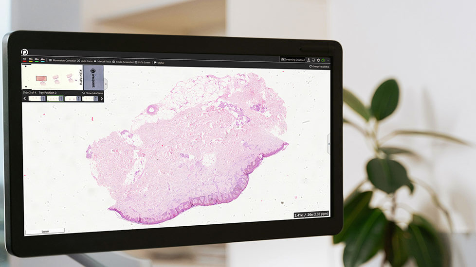
In the digital pathology environment, the whole slide imaging (WSI) technique is one of the most important aspects of modern pathology practice. Digital pathology has created an environment conducive to the seamless utilization of WSI. This technology enables effortless implementation of methods like image visualization, integration of artificial intelligence, and digital scanning, thereby enhancing the overall application of WSI. With the whole slide imaging, there is no threat of breakage or damage to the staining of the physical slides. PreciPoint’s Fritz Microscopy Slide Scanner, your digitization helper in microscopy, can effortlessly digitize histological sections in first-class image quality. It offers you the possibility to view high-resolution images by using all the touch-enabled devices you have at your disposal. Furthermore, the iO:M8 Digital Live Microscope, M8 Microscope and Scanner, and O8 Oil Digital Microscope and Slide Scanner serve to enhance the advantages of working with WSI in the context of patient treatment. These advanced devices facilitate seamless navigation through microscope slides and enable the necessary observations to be made with greater ease and precision.