
All intraoperative procedures have one thing in common: time. Time is what matters first and foremost. How quickly can the frozen section report be communicated back to the surgeon?
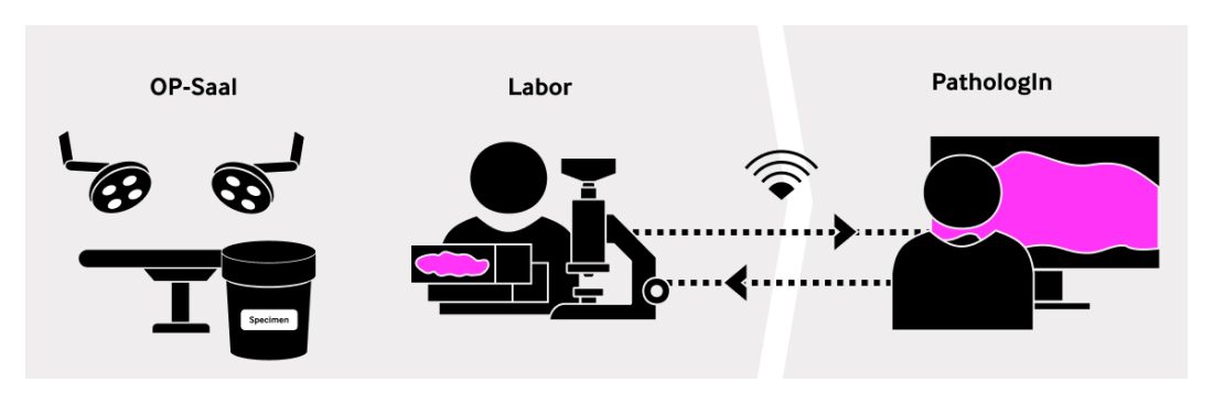
From a regulatory perspective, the report for a single intraoperative frozen section slide must be delivered to the operating surgeon within 15 minutes (20 minutes in some countries) after the surgical specimen has entered the lab. During the consultation, the specimen is dissected and processed as it moves through macroscopy and cryosection through to the rapid microscopic evaluation.
There is no time to lose, everything must run like clockwork.
The Digitization Challenge of Intraoperative Consultations
In any field of work, the adoption of new technology is always driven by the need to reduce downtime, to improve efficiency, productivity, and quality, or to automate tedious and repetitive tasks. In the end, we all want to achieve more with less effort, so we can spend time doing other important things.
Digitization of intraoperative consultations can offer many benefits to pathologists, surgeons, as well as patients. Probably the most compelling benefit of digital technology is the fact that digital technology enables pathologists to refer a case to a specialist or sub-specialist for an immediate second opinion. Not only does it enable a rapid second opinion in the first place, but it also allows to tap into a much broader pool of specialists way beyond regional borders. More benefits are outlined in our article here: Click here to find out what are the 3 reasons why pathologists may seriously consider digitizing intraoperative consultations.
Given the significant benefits of digitization, the main question for pathologists is to understand which technology to use. What is the best way to digitize the traditional, analogue microscope?
Stay Ahead with Insights from Precipoint!
Welcome to our newsletter! Be the first to know about our latest products, services, webinars, and happenings in PreciPoint. Don't miss out on this opportunity to stay informed. Subscribe to our newsletter today!
By clicking “Subscribe”, you agree to our privacy policy.
Is digital pathology suitable for intraoperative procedures?
Definition and Scope of Digital Pathology
Digital pathology is commonly defined as a sub-field of pathology that focuses on data management based on information generated from digitized specimen slides1. So called Whole Slide Images (WSI) are the output of scanning devices that generate digital representations of physical specimen on glass slides. In the rest of the article, I refer to that technology and process interchangeably as digital pathology, WSI, or scanner.
Digitization in Routine Pathology vs. Intraoperative Pathology
Much of pathology research has already been digitized over the last 10 years. As of lately, many healthcare centers, hospitals, pathology institutes and laboratories have started the process of digitizing clinical workflows (also referred to as routine pathology), where WSI shall be at the core of all processes and communications. It is therefore very tempting to think that WSI are the answer to the intraoperative digitization challenge. But are they really? Can we transfer what we are implementing in routine pathology for use in intraoperative microscopic assessment during surgical pathology?
Limitations of Scanning Technology in Intraoperative Pathology
The technology used for scanning routine pathology slides and especially scanners have been optimized for fast throughput and for samples used in clinical pathology workflows. It works best for standard high-quality formalin-fixed, paraffin-embedded (FFPE) slides with no or very little artifacts.
Challenges with Scanning Complex Slides
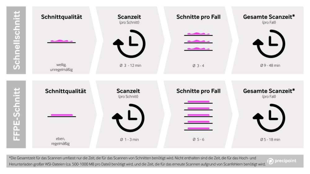
Scanning uneven slides or slides with cutting or ice crystal artifacts as found in intraoperative consultations don’t deliver the same quality image within the same time frame as routine slides. It takes significantly longer to scan complex slides because other scanning settings must be used and the output may still be a poor digital image quality. For more details, you may refer to our Surgical Pathology page.
Increased Scanning Time for Non-Standard Slides
According to our research, scanning time between FFPE and frozen section slides can be multiplied by four. What takes a couple of minutes for a standard FFPE slide may take up to 10 minutes for a complex slide such as a slightly fatty cryosection. With a little hindsight, pathologists working with WSI-based digital pathology agree that a major constraint is the scanning time, especially with non-standard slides that differ significantly from common FFPE slides coming out of a routine histology lab.
Multiplicity of Slides in Frozen Section Cases
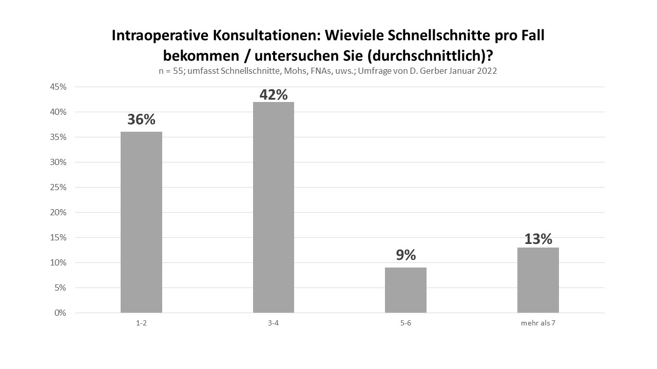
What is more, to consider is that frozen section cases usually consist of more than one slide. A survey conducted in January 2022 with 55 pathologists showed that 64% handle more than 3 slides per case, with 42% having an average of 3-4 slides per case. Therefore, you need to multiply the total scan time by the number of slides per case. As a result, in most cases, the slide scanning process alone will end up taking longer than the 15 minutes prescribed for the whole consultation. That renders the use of such technology prohibitive.
Quality and Technical Challenges with Scanning Frozen Section Slides
A main issue is the need to rescan a slide when the quality of the WSI is insufficient for diagnostics. This concerns difficult, low-quality slides which is exactly what frozen section slides are. The underlying reason is, as described above, the sample preparation methods specific for intraoperative consultations. Some studies found that up to 13% of cases produced WSI could not be used for diagnostics2. That number is unsustainably high in a productive work environment.
Challenges with Wet Mounting Medium and Closed-Box Scanners
Another issue pathologists and lab technicians have reported is that wet mounting medium as commonly found in frozen sections may drip off glass slides during use. That is a major problem when operating scanners that are designed as so-called closed-box instrument. Users might not be able to clean such instruments, which will over time probably result in severe functional problems of the device.
Suitability of Scanners for Intraoperative Consultations
Prospective users should also investigate whether the device is actually intended for use for the purpose of a rapid microscopic evaluation of surgical specimens during intraoperative consultations. Often, scanners are intended to image histology slides prepared from formalin-fixed, paraffin-embedded (FFPE) tissue. Whole slide imaging systems may not be intended for use with slides prepared from frozen tissue, cytology, or non-FFPE hematopathology specimens.
Exploring Telemicroscopy as an Alternative
Considering Other Technologies for Intraoperative Consultations
Here is the bottom line. What works for research or routine may not be the best solution for intraoperative consultations. In other words, intraoperative processes cannot be compared to routine processes. Seeing that slide scanners may not actually present any significant time benefits for intraoperative consultations, it is worth looking at other technologies, such as telemicroscopy – also known as digital microscopy – as an alternative and potentially more practical solution.
Telemicroscopy for Hands-On Teleconsultations
Telemicroscopy resembles the manual analog microscopes and places the pathologist at the center of the process.
Instead of scanning the frozen section glass slides, the pathologist or laboratory assistant places the frozen section glass slides under a digital microscope. The pathologist can now immediately assess the slides on his screen at the point-of-care – or remotely – since the displayed image is a streamed microscopy image. If the pathologist is working from a remote location or at home, a microscopy streaming software allows them to access the digital microscope from their remote workstation.
What´s more, with a fully motorized digital microscope (x-y-stage, z-axis), the pathologist is also able to fully control and steer the microscopy hardware remotely: at the move of a fingertip on a display or mouse, the user can navigate, zoom in, and refocus as if they were using the microscope at point-of-care.
Overall, the use of telemicroscopy technology empowers pathologists to have full control over the observation of the slide remotely, in real-time, and digitally.
The Data Challenge
Whole slide images produced by scanners are permanent image files and often include patient data. They are extremely large, usually between 400 MB to 2 GB per image file. The management and distribution of these heavy data sets is challenging, especially within the limited time frame of an intraoperative consultation and within commonly existing IT infrastructure that was not designed to handle such huge data on short notice.
A telemicroscopy solution brings superior advantages and adds a lot of flexibility. Unlike WSI-based digital pathology, the assessment of live microscopy images on a screen makes the storage and transfer of large WSI files fully redundant. Telemicroscopy doesn’t require any permanent storage or server space, as the live microscopy images are only temporarily cached (stored) on the computer and no longer available when the application is closed. When the consultation needs to be held remotely, a streaming connection to the microscopy control software can be established within the network of the healthcare institute or across different networks through fast enough Internet connection – which is mostly available.
Conclusion - More flexibility with telemicroscopy
(2) Source: Intraoperative frozen section consultation by remote whole-slide imaging analysis
–validation and comparison to robotic remote microscopy
Thomas Menter, Stefan Nicolet, Daniel Baumhoer, Markus Tolnay, and Alexandar Tzankov