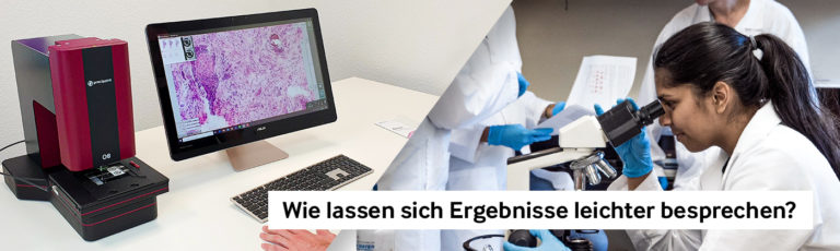

Benefits of Digital Microscopy in Education
Enhanced Teaching Experience with Digital Microscopy
Since the conventional microscope is not ideally suited for educational use, students and teachers alike benefit from digital microscopy. It allows everyone involved to examine and discuss the same sample. In this blog post, learn how far this technology is helping educational institutions move forward.
Stay Ahead with Insights from Precipoint!
Welcome to our newsletter! Be the first to know about our latest products, services, webinars, and happenings in PreciPoint. Don't miss out on this opportunity to stay informed. Subscribe to our newsletter today!
By clicking “Subscribe”, you agree to our privacy policy.
Digital microscopy promises many benefits for both students and educators. If teachers or educators have the right tools, teaching can become an experience for all. Previously, teaching was done using traditional light microscopes. Lecturers would give their lectures on scientific topics and students would then view the specimens through their individual microscopes. [1] This approach inherently presented some problems.
Overcoming Challenges of Traditional Microscopy
For example, students could only look through the eyepiece one at a time and had to make room for the instructor to show them specific locations in the specimen. This way of working naturally made communication difficult. By contrast, when the entire course can access and navigate a digital image, the result is a collaborative, rounded class.
Promoting Teaching and Communication with Digital Microscopy
Advantages of Shared Virtual Samples
The teaching of the content and communication is promoted by:
- Everyone can view the same virtual rehearsal and therefore talk about the same content
- The same important sections and “events” on the sample are visible to everyone
- Students focus on the lecture instead of frantically looking into their eyepieces
- The instructor can show students key details in the presentation
Accessibility and Cost-Efficiency
So that every student or trainee has access to the same sample, there is no need to buy a separate digital microscope for everyone.
The scanned samples are simply opened on the notebook via the viewer software and processed from there.
Digital microscopy therefore holds immense potential. Students can work together without having to take turns at the microscope, and instructors can discuss and show results as a group. By comparison, conventional microscopes lag in handling. They offer far less interactivity and flexibility.
[1] Sagun, L., & Arias, R. (2018). Digitale Pathologie: Ein innovativer Ansatz in der medizinischen Ausbildung. Philippinische Zeitschrift für Pathologie, 3 (2), 7-11. doi: 10.21141/pjp.2018.009