
Synopsis
The rising number of cancer cases coupled with a declining pathologist workforce poses significant challenges in cancer diagnostics. This trend necessitates innovative approaches and streamlined processes in pathology to ensure timely and accurate diagnoses for cancer patients. Addressing this issue is crucial to meet the growing demand for pathology services and optimize patient care in the face of increasing cancer prevalence. Digital microscopes such as the iO:M8 by PreciPoint and the digitization of laboratory workflows in surgical and clinical cancer diagnostics help pathologists to work more productively and efficiently. How? Here are the advantages of using a digital microscope in pathology.
Role of a Pathologist
The job of a pathologist is to identify abnormal changes in tissue or cell structures and then match those with disease patterns. The first step involved the pathologist examining the surgically removed tissue sample macroscopically, i.e., with his bare eyes. Then he microscopically analyses the tissue section produced from the specimen to describe it in detail and identify the changes. As evident, the microscope is the most essential working tool for analysis.
What Distinguishes Digital Microscopes from Analogue Microscopes?
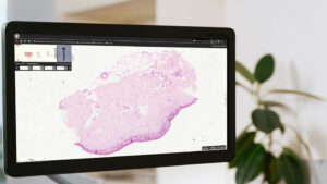
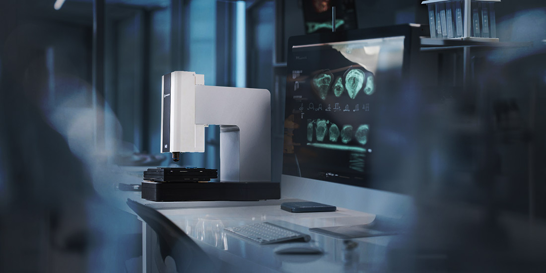
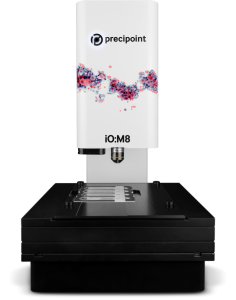
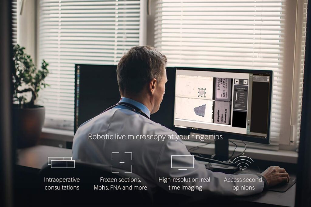
Digital microscopes such as the iO:M8 by PreciPoint have a camera instead of an ocular and the tissue sample is fixed in a slide holder. The image of the tissue section is transferred to a computer and displayed on a high-resolution screen. The pathologist can now control the digital microscope on the screen, navigate over the tissue section at the click of a mouse and examine the specimen in real-time. As a result, digital microscopes offer significant advantages.
What Are the Advantages of Digital Microscopes When Examining Specimens?
Digital microscopy contributes to ensuring the quality of examining specimens. It also facilitates the work and everyday life of pathologists thanks to the following plus points.
#1 High-resolution Image Quality
Digital microscopes produce high-quality, high-resolution images in real-time. They deliver high-precision images of not only simple tissues but even of complex specimens such as smears or frozen sections, for rapid microscopic assessment on screen.
#2 Comprehensive Views
Digital images of specimens can be greatly enlarged and viewed in detail. Users can quickly and easily navigate to a region of interest in the live image, zoom in, and seamlessly refocus to the desired details on the screen. The sample can also be analyzed from different angles on the screen.
Digital microscopes often come with multiple programs, e.g., functions to increase contrast, add inscriptions and annotations, and much more.
#3 Speed
Digital microscopes accelerate the whole process. The slide can be quickly mounted on the holder of the digital microscope and the specimen is immediately available for microscopic examination on the screen. There is no wait time and no need to create whole slide images by scanning the slides.
#4 Improved Ergonomics
Digital microscopy on the screen can be performed comfortably in an upright position, making it more convenient and ergonomic than looking through an ocular. A healthy sitting position for the user prevents incorrect physical strain and the resulting damage to health. This in turn increases work satisfaction and productivity.
#5 Flexible Working Concepts
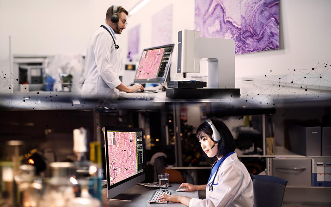
Digitized slides enable on-screen evaluation even from home. This opens the door for flexible working concepts in pathology – another advantage in view of the acute shortage of pathological experts.
Digital live microscopy has considerable advantages, especially when, for example, in difficult cases, consultations between remote experts and a rapid interdisciplinary exchange are required.
How Does Live Microscopy Simplify the Exchange between Experts?
Digital microscopes expedite communication and allow a way to obtain second opinions quickly and easily. Better exchange of knowledge benefits pathologists and physicians by not only improving the quality of diagnosis and treatment but also increasing access to remote expertise.
Microscopic Examination from a Distance
Pathologists can make the images of a specimen available to other experts or grant them access to their own computer via streaming software when using a digital microscope. This remote analytical capability allows easy and fast collaboration between specialists across multiple locations, national borders, or even continents.
Second Opinion without Delay
The images can easily be made available digitally for immediate assessment by other specialists. Obtaining a second opinion is thus possible within a very short time without any delay due to transport or travel times.
Using the PreciPoint Streaming Software, you can easily grant remote specialists access to your computer and thus obtain a second opinion in the shortest possible time. This makes the iO:M8 as well as other digital microscopes particularly suitable for use in time-critical intra-operative examinations and consultations.
Digital microscopes such as the iO:M8 by PreciPoint score with high image quality, detailed views and a computer workstation that makes the daily routine of pathologists more ergonomic and flexible. They may facilitate professional exchange between experts at different locations. This increases efficiency in pathology laboratories as well as quality in cancer diagnostics.
Conclusion
The increasing incidence of cancer, coupled with a declining pathologist workforce, poses significant challenges in cancer diagnostics. To address this issue, novel approaches are essential. Digital microscopes like the iO:M8 by PreciPoint, combined with the digitization of laboratory workflows in surgical and clinical cancer diagnostics, offer a solution. iO:M8 empowers pathologists to work more efficiently and productively. They enable the rapid examination of tissue samples, seamless sharing of results, and the potential for remote consultations. This not only helps manage the growing workload but also ensures timely and accurate cancer diagnoses, ultimately improving patient outcomes.