
Synopsis
Scientists, students, trainees, researchers, and pathologists work with digital microscopes daily. Thus, the right functioning of the microscope is imperative. For a digital microscope to work at its optimal best, it is important that it is taken care of. Proper functioning and achieving the right image clarity, magnification, lighting, and depth of field are key to an accurate diagnosis. Using up-to-date software and proper cleaning and maintenance should be done on a regular basis.Appropriate steps must be taken on time to mitigate any issues arising from the functioning of the digital microscope. To achieve this, it is critical to collaborate with a reliable partner who can offer digital microscopes of the best quality and services at affordable prices. This article will delve into the importance of a digital microscopes’ proper functioning.
Digital Microscope Function
In today’s world, digital microscopes are used in a variety of fields. In scientific research, experts use it to study and analyze a wide range of specimens. Microorganisms, cells, bacteria, minerals, and other materials can be easily observed and studied under a microscope. Many students and trainees use digital microscopes for education and training, where they can study specimens at high magnifications without the need for slides and lenses used with an analog microscope. Industry and the manufacturing segment also use digital microscopes for quality control, inspection, analysis, and quality evaluation.
Multiple uses of digital microscopes
… for viewing and analyzing biological samples, tissues, and cells.
… for conducting scientific studies and experiments.
… for teaching students about different specimens and objects.
… for examining and evaluating industrial components, surfaces, and materials.
… for inspecting products and ensuring they meet quality standards.
The Role of Digital Microscopes in Modern Medicine and Pathology
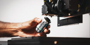
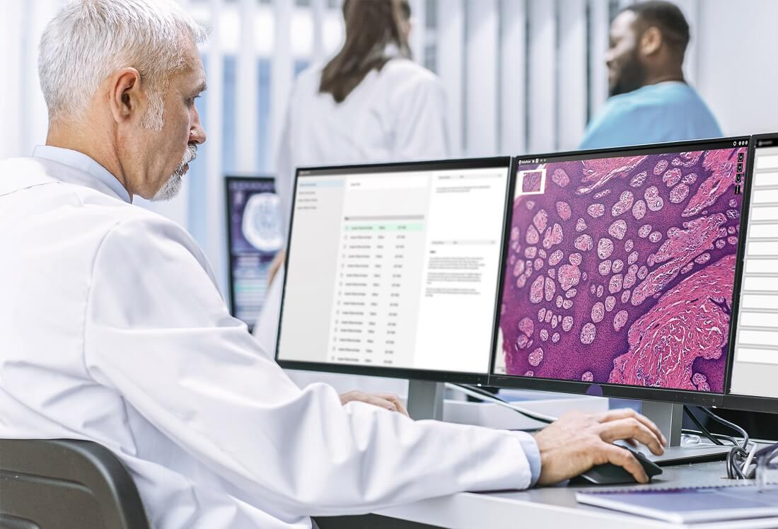
A digital microscope is an integral part of modern medicine. Digital microscopes make the process coherent, especially when it comes to pathology and surgery. In pathology, a digital microscope is used, especially in diagnostics, to observe the characteristics of the disease. The hardware of the digital microscope consists of an analog microscope, a light source, a camera, and the camera housing. The camera replaces the position of the user’s eye. The image of the observed object is focused on the camera and it is continuously displayed on the user’s computer screen. The user captures the image of the observed object by selecting the appropriate menu on the computer screen. Once the image is captured, it can be stored or processed depending on the user’s need.
Digital Microscope Parts and Functions: The Importance of Proper Working
The possibility to observe fine details and provide accurate and detailed information about the specimen is one of the main reasons why a digital microscope is a critical tool. To achieve the best results, digital microscope parts need to function properly. The entire research and analysis can have an impact on the conclusion, outcome of the diagnosis, and other phases in the prognosis and research.
Lives at Stake
If something in the management of the data obtained by the digital microscope goes wrong, it can seriously affect the final results and even jeopardize the further workflow. Proper functioning of digital microscopes is critical, especially in medicine and healthcare, because one incorrect observation can affect the entire diagnosis and, ultimately, the quality of care and treatment and even threaten the life of the patients. A properly working digital microscope is crucial to avoid misdiagnosis and mistreatment.
Steps for optimal use of digital microscopes
Properly assemble and securely mounted the microscope to a stable surface.
Lighting is sufficient and properly adjusted for optimal viewing.
Use the appropriate lens for the sample and adjust the focus accordingly.
Prepare the sample for optimal viewing and analysis.
Use the appropriate software and storage method for capturing and saving images.
Regularly clean the microscope and its components.
Be familiar with common issues and their solutions.
Educate all users on the use of the microscope and its software.
Compromised Output
Researchers and other medical experts, often use a digital microscope for research purposes. If a digital microscope or some parts of a digital microscope are not functioning properly, then the results obtained lead to the wrong conclusions that result in wasted resources and failed research. For example, incorrectly performed tumor cell research results in incorrectly set diagnoses. Inadequate microorganism research results in inaccurate data, etc.
Basic Requirements
For the best functionality, working conditions must meet two basic requirements: (1) a user interface to allow humans to enter information and (2) a mechanism to convey this information back into the workflow environment. To ensure everything is working properly, it is necessary to incorporate human feedback within workflows. Human decision-making capacity is an aspect that cannot be neglected.
Solutions to Overcome the Challenges
Medical organizations and institutions can encounter some common function limitations or issues when it comes to working with digital microscopes. The first issue could be image clarity. An image obtained from a digital microscope needs to be clear and in high resolution. Also, poor lighting can often produce blurry and grainy images. If your work with certain tissue specimens, you need to pay special attention to light adjustment because sometimes a digital microscope can have limited possibilities of light adjustments. Magnification can be crucial when working with tissue samples, so you need to be familiar with the magnification possibilities of the digital microscope. Some digital microscopes can also have limitations regarding the depth of field, so attention also needs to be paid to these things. To overcome the issue, you can look at replacing the objective lens with a higher magnification lens or increasing the size of the camera sensor to capture more detail.
Effective approaches to overcome the challenges
Familiarity with the Software
Another concern could be with the software. If the user is unfamiliar with the software, it can be challenging to handle the whole workflow. Thus, it is always ideal to educate the user on the features of the microscope and train them to operate the microscope properly. Sometimes, a digital microscope may be unable to connect to the software. The reasons vary from computer to computer. Plugins, viruses, memory capacity, and even hardware problems can limit the work output. Some digital microscopes aren’t compatible with certain computer systems or software. You can overcome such problems by updating the microscope software to the latest version which would include fixing bugs and improving performance. You must also check the computer’s compatibility with the microscope software to ensure it meets the minimum system requirements and that it is configured properly.
Patience Pays Off
It is also worth mentioning that sometimes too much cleaning and maintenance can limit the user from achieving the best possible results. A digital microscope is a complex tool that requires certain amounts of time to do cleaning and maintenance. If that’s not organized properly, working efficiency can be decreased.
What Needs to be Done?
Laboratories and institutions that work with digital microscopes need to ensure proper maintenance of the digital microscopes and ensure that they always work properly. It is necessary to collaborate with a reliable client or partner who can offer digital microscopes of the best quality. That doesn’t only mean the best hardware and software, but also the best service. High-quality manufacturers need to be available so that customers can always contact them in case there are issues.
Conclusion
Digital microscopes are used daily by a variety of professionals, including scientists, students, trainees, researchers, and pathologists. Ensuring that these microscopes are functioning properly is essential for accurate diagnoses. Achieving the right image clarity, magnification, lighting, and depth of field is key to achieving this accuracy. To achieve optimal performance from digital microscopes, regular cleaning and maintenance, as well as the use of up-to-date software, are necessary. Prompt action must be taken to address any issues that may arise from the functioning of the microscope. To achieve this, it is important to work with a trustworthy partner who can provide high-quality digital microscopes and services at reasonable prices. While PreciPoint offers fast turn-around technical support, its fully motorized live microscope for rapid on-site evaluation iO:M8 is designed as an open platform for easy access and cleaning.