
Synopsis
When observing modern digital pathology practice, especially in science and education, digital microscopes are the real game changers. The question now arises is how? There are two main reasons. The first is the numerous features and benefits that digital microscopes have, and the second are the opportunities that they offer. Features like magnification, high image resolution, and other advanced image possibilities open doors to a new knowledge base, detailed information, accurate diagnosis, efficient treatment, and the unique learning experience needed in the modern scientific environment. All these aspects and their importance for modern science and education will be discussed in the following text.
Are Digital Microscopes Any Good?
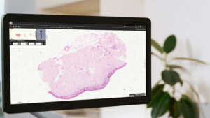
One of the fundamentals of every science and education program is the use of a digital microscope. While traditional microscopy held an important place for a long time, the high costs of maintenance made scientists search for an alternative. Digital microscopy was the solution to this search of theirs. In digital microscopes, slides embedded with blood specimens are converted into a digital form that can be run with computer-driven software. In education, a digital microscope is an effective tool, just as it is in science, in a medical laboratory.
Supporting Advanced Technology in Science
When it comes to science, a digital microscope completely changes the game. Since the introduction of charge-coupled device (CCD) cameras for image acquisition through the microscope in the late 1990s, scientists have been working on assembling a series of digital microscopic images into giant montages for making whole microscopic slides accessible through the computer monitor.
Major Drawback Addressed
In the early 2000s, digital microscopy had one major flaw. It lacked the proper IT support for handling the thousands of MB representing digital microscopic slides. Those were the early days of this practice. IT technology has taken giant leaps compared to the early 2000s. It is now easy to handle the large amount of data running into GB. The revolution is believed to continue in the coming 5-10 years in all fields including science like its use in histopathology.
Accurate and Efficient Research
Scientists have an integrative role. That means, by using histopathology, they can diagnose malignant tumors and screen for biomarkers related to patients’ responses to molecularly targeted therapy. The digital microscope allows a better understanding of the biological backgrounds of disease processes, tissues, and cell morphology. With a digital microscope, it is possible to apply certain techniques of molecular morphology that involve combining structural and molecular information. This method helps in detecting chromosome and gene abnormalities, immunohistochemistry (IHC), and reveals translated proteins in normal and diseased cells.
Additional Power
A digital microscope also helps to determine semi-quantitatively a series of biomarkers at the cellular or subcellular level within the tissue architecture. A digital microscope offers unique features that are not available in conventional optical microscopy. Assisted by dedicated software tools, it permits dynamic and prompt access to stained slides at multiple microscopic magnifications as controlled with a mouse through the computer monitor.
Raising Standards
The calibrated qualities of discrete pixels raise the level of automation of image analysis and quantification and make the diagnostics or research work easier and more efficient. The unlimited access to slides makes a digital microscope a very efficient tool for telepathology, diverse types of diagnostics, teleconsultations, proficiency testing, external quality assurance, and interlaboratory process validation.
Opportunities in Education
One of the biggest advantages of the digital microscope is its versatility and reasonable cost. The benefit of its low cost is evident when one considers the burden of purchasing and maintaining optical microscopes and the regular replacement of tutorial glass slides. In education, a digital microscope brings the advantage of accessibility to everyone, or, in other words, the best selection of standard slides without spatial or temporal limitations. A single digital slide can demonstrate a rare entity or any expensive ancillary technique such as IHC to an unlimited number of students.
Flexibility for Students
The students can view two or more slides in parallel, open, and correlate several copies of the same slide, or follow annotation labels and text adapted for distant learning. Digital images offer the means for extending and building upon traditional methods of inquiry. By capturing the things that wouldn’t be observable without the digital microscope, the students can share conclusions about certain subjects. Along with science, the digital microscope changes the game in education because it represents a new way to gain new knowledge and new skills crucial for students and future experts in the new era.
Numerous Advantages
A digital microscope represents a high-quality solution for the contemporary research lab or the classroom. It is an advanced technology with a range of benefits that involve higher magnification, improved imaging resolution, and advanced image processing tools. Digital microscopes enhance the opportunity to acquire new knowledge, make conclusions in research, or learn with greater efficiency. It is a crucial tool for scientists, educators, and diverse enthusiasts. It allows for discoveries and insights in a variety of fields.
Conclusion
Digital microscopes have significantly changed the technical working environment of researchers and students. Compared to traditional optical microscopes, digital microscopes including our fully motorized digital live microscopes offer a range of benefits like magnification, improved image resolution, and advanced image processing tools. These factors lead to a better understanding of the research in a more advanced way that provides greater accuracy and precision. In education, the digital microscope is an asset because it provides a unique experience for knowledge gain for students and experience gain for professors. All-in-one virtual microscopy platforms such as PreciCloud make the whole process of teaching and learning more efficient and accurate. It provides new expertise for students as it allows them to access in-depth knowledge, which is necessary in the world of digital pathology, which is constantly changing and improving.