
The field of pathology is going through a digital revolution. Digital technology has opened new avenues for students and practitioners to learn and improve their skills. One of the critical advantages of digitization in the field of education is standardized course material. It also offers pathologists and students access to endless amounts of images, without the need to travel to another location or country to attend an event. Virtual opportunities enable them to learn and expand their knowledge base conveniently and consistently.
Digital Revolution
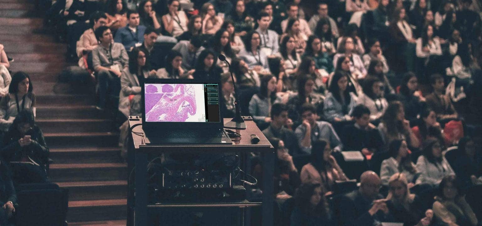
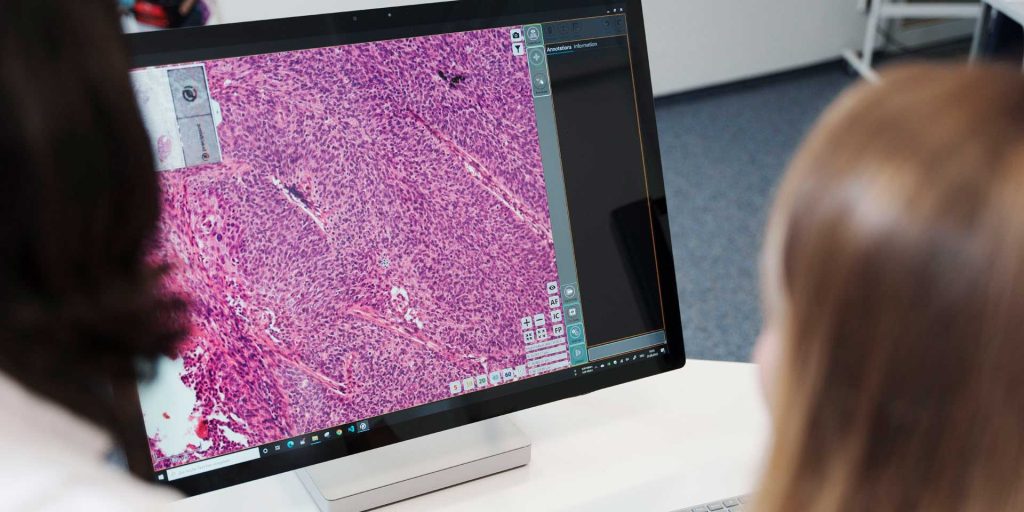
The field of pathology is going through a digital revolution. The rapid transition from traditional light microscopy to digital microscopy is greatly benefiting the education sector. The times are changing, and with them, technology is advancing. With the emergence of whole slide imaging (WSI) technology paired with other web-based pathology resources, pathology, and medical students are experiencing a whole new way of learning. Digital technology in pathology education is empowering students to pick up skills that they can effectively apply in a real pathological environment. They can now easily identify areas of diagnostic relevance from an entire slide by simply using a computer.
How Does Digital Technology Help in Teaching?
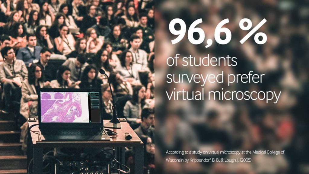
A survey conducted by the Medical College of Wisconsin (MCW) highlighted some valuable insights. First-year medical students (M1s) who used the virtual microscope for histology laboratories in 2004 and neuro courses were asked to subjectively estimate the overall effectiveness of the virtual microscope for learning. 98.5% rated the effectiveness of the virtual microscope for learning as excellent or good. 96.6% of the students agreed or strongly preferred to learn via the virtual microscope over the light microscope.
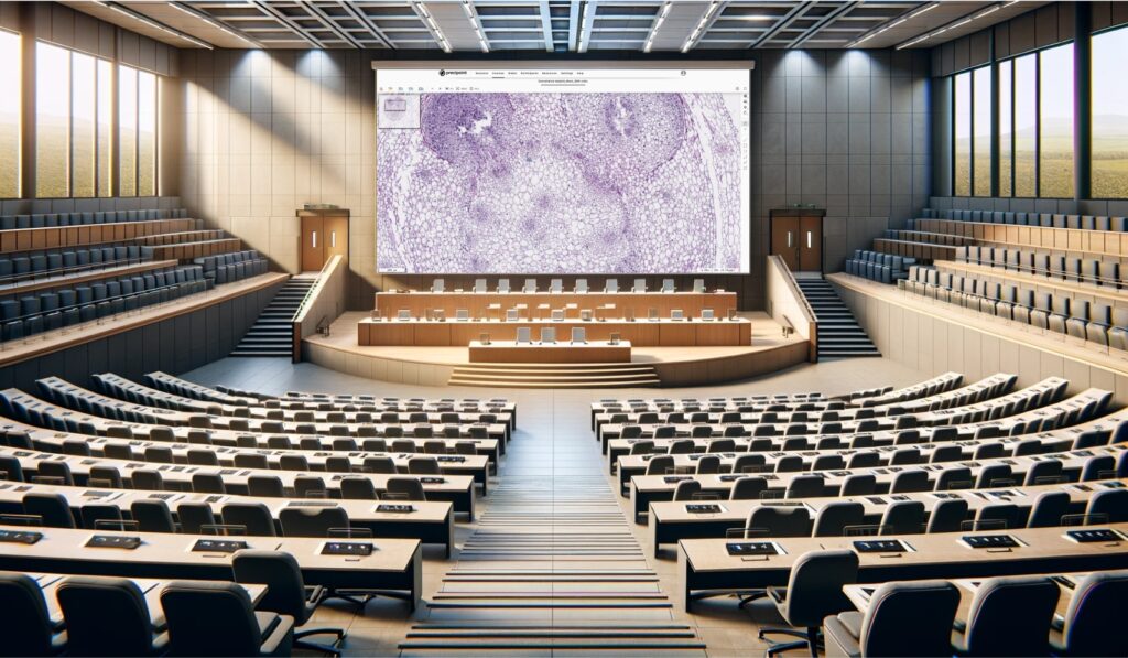
With the help of digital pathology, course material can become standardized. With light microscopy, students usually had a microscope to view histological slides. The problem was that they were not always looking at the same cut. Some students may not see the same morphological and biomarker expression patterns. With digital pathology, all the students see the same sample. There are no learning discrepancies. Each student has the same learning opportunities and quality of education. It is understood that standardized course material produces a better quality of learning.
Stay Ahead with Insights from Precipoint!
Welcome to our newsletter! Be the first to know about our latest products, services, webinars, and happenings in PreciPoint. Don't miss out on this opportunity to stay informed. Subscribe to our newsletter today!
By clicking “Subscribe”, you agree to our privacy policy.
Role in Education
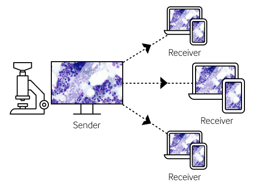
We are seeing a rising trend in the implementation of digital pathology technology in the education sector in many countries. There are even some countries that are using telepathology in education. The opportunities that arise with telepathology technology are endless. If we take medical congresses as an example, there are numerous virtual opportunities where attendees don’t have to travel to a different country or location to attend the event, and they have continuous access to endless amounts of images.
Implementation Challenges in Telepathology
Telepathology can be used in the continuation of medical education from undergraduates to postgraduates, each using it a little differently. The result is still the same; they can transmit digital images to enhance their learning. The implementation is in full effect, and if this implementation trend continues, virtual microscopy could make a traditional microscope a thing of the past. However, it is not always that easy to implement these changes. Things might seem easy, but in fact, some challenges must be addressed.
Infrastructure and Labeling Considerations
The implementation of digital microscopy in medical education does not pose the same challenges as it would in clinical diagnostic practices. There are, however, some things that must be considered. There must be a system in place for correctly labeling slides so that they can be accurately identified in the database. With new technology comes new infrastructure that allows images to be transferred. The creation of a digital microscopy lab is an initially expensive project. However, it may become more cost-efficient in the long run. Dee et al. calculated the cost of a microscope laboratory for 50 students to be about $100,000 per year, which approaches the complete start-up cost for virtual microscopy, including the purchase of a virtual slide scanner.
Internet Accessibility and Technical Issues
Aside from the initial cost of the infrastructure, the next challenge is the internet. We must remember that in this world, not everyone has access to the internet. Undeveloped or low-resource areas may not have the same luxuries as developed areas. On top of that, academic networks are often slow and expensive. Within the academic setting, students must be able to access computer workstations, especially after class hours. With technology, there is always something that can go wrong. It is not perfect, and even the slightest problems electrically can cause an outage. For example, if the weather were bad, it might disrupt telecommunications.
Impact on Student-Teacher Interaction
Lastly, there is a concern about whether student-teacher interaction would dynamically decrease due to the implementation of digital microscopy. The truth of the matter is that this technology enables students to learn pathology in a more communal and stimulating manner.
PreciCourse: Digital Learning Solution
A premier destination for education in virtual microscopy and quality management.
- Interactive Digital Courses
- Advanced Slide Viewing
- Comprehensive Evaluation
Opportunities for Growth
Enhancing Pathology and Life Sciences
With the aid of technology, digital microscopy and, more specifically, digital pathology, have so much growth potential. The growth potential is not just confined to pathology; other life science disciplines can also reap the benefits of digital microscopy. Decision support, conferencing, proficiency testing, and quality assurance can all benefit from digital pathology. Unlike reference books, relevant details in a digital slide can be viewed “in context” to provide pathologists and students with comprehensive, evidence-based pathology content to facilitate self-teaching or decrease the time required by pathologists to research complex pathology cases. Over the course of their careers, many pathologists assimilate collections of glass slides that they sometimes use as reference slides for decision support. Digital pathology makes it possible for pathologists and medical students to access digital slides from anywhere at any time.
Quality Control and Training
Digital pathology can also be deployed in training and quality control practices. In the past, quality control was done by sending physical glass slides to other external partners to look at and review. The cost of sending glass slides externally makes it much more difficult to provide quality control. Digital microscopy makes it simple to make digital slides accessible to other institutions. Digital pathology has the potential to create a database for organizations to use. The sharing of digital images has never been easier. Like quality control, practical training is another customer for digital pathology.
European Initiative for Competency Tests
Trainees or residents in the field of pathology can also benefit from digital pathology. It all revolves around the ability to access an entire histological slide to understand the pathological context. The European Association of Pathology Chairs and Residency Program (EAPCP) has started a major project throughout Europe using online competency tests for residents in pathology. It is an excellent way to train these residents who are coming up in the pathology world. The way the competency tests work is that there is a mixture of questions, all based on digital slides. The residents can view these slides virtually and answer questions based on the material. Then the residents can compare their performance with that of their peers. The EAPCP has been able to effectively implement competency tests for training in 27 countries in Europe.
Market Growth and Future Prospects
From teaching to training, digital technology will not die right now but will grow in the future. According to Allied Market Research, “the digital pathology market accounted for $512 million in 2018 and is expected to reach 1,390 million by 2026, registering a CAGR of 13.3% from 2019 to 2026.” The market is growing because technology and the need for technology are growing. One digital device can do the work of five different older devices that pathologists used to use. The major factors that are pushing the growth of digital pathology are the cost efficiency of digital pathology products, how easy it is to transport virtual slides, and the high efficiency of digital pathology systems. In general, there is a big push for digital pathology, and there are several applications that can adopt the new technology. That creates new demand for digital pathology in all the different applications.
Connect with our PreciCourse Experts
Interested in learning more about PreciCourse or have specific inquiries? We’re here to help. Use the form below to get in touch with our experts.
Your privacy is important to us. PreciPoint uses your information to contact you about relevant content, products and services.
You can unsubscribe from any communication at any time via the footer of our emails. You can find more information in our data privacy policy.
Conclusion
Digital technology is providing students and practitioners with new channels to enhance their skills and knowledge. A key benefit of this transformation is the uniformity of course materials, which helps to ensure consistent quality across different learning platforms. In addition, digitization offers pathologists and students unparalleled access to an unlimited amount of image data, without requiring them to travel to distant locations to attend events. This convenience and consistency enable them to learn and expand their expertise conveniently and flexibly. Digital microscopes from PreciPoint make teaching across universities around the globe consistent and systematized. Moreover, the virtual microscopy platform PreciCloud allows setting up seminars for advanced training and lets users create online courses with virtual slides.