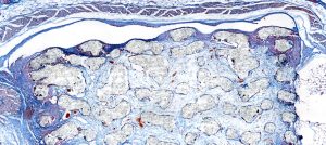

Sandra Haßelt views histopathological slides in orthopedics and view scores. For example, Brackertz score (Arthritis score) and Krenn score (Synovitis score) to find out changes in the synovial layer, like infiltrates of macrophages or hyperplasia. We also see changes in ossification and cartilage.
Challenges to be solved:
We used to use a traditional microscope for our research. When using a traditional microscope, we don’t have a large overview of the specimen, and the scope of the field is very small. Therefore, the tissues and specimens that we need to view are too big for our little microscope. It took too much time to evaluate the scores and conduct statistics. We sometimes have 600-800 slides to look through so that we can count specific cells and immunostained cells on each slide. The countless hours it takes to look through all the slides though a traditional microscope was unbearable. With our previous microscope, we would have to take 20 separate images to view the entire specimen. Then comes the problem with manually stitching the images together into one large image.
Stay Ahead with Insights from Precipoint!
Welcome to our newsletter! Be the first to know about our latest products, services, webinars, and happenings in PreciPoint. Don't miss out on this opportunity to stay informed. Subscribe to our newsletter today!
By clicking “Subscribe”, you agree to our privacy policy.
“Fast evaluation. Brilliant pictures with high resolution. Easy and simple handling.”

Sandra Haßelt
Medical Technical Laboratory Assistant at Ludwig Maximilian University of Munich.
Our approach and solution:
The M8 has completely changed our workflow process. We now have a large field of view where we can see the whole specimen. There is no need to take 20 different images and try to manually stitch them together. The M8 automatically stitches the images together for us in real-time with the same illumination. Now it’s very easy. We have a big overview of the entire slide immediately and can zoom into any part without switching objectives. Also, the stepless zoom allows us to have a view of the specimen at “in-between” magnification. Using the M8’s unique mode, instant scan, we can deal with a high number of slides 3-4 times faster than using a normal microscope. The instant scan mode also allows us to annotate. The line tool allows us to measure the distance between the cartilage and the synovial layer. We also use the counting tool to count cells that are colored in immunohistochemical staining.