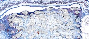

Dr. habil. Tatiana Flisikowska works at the Technical University of Munich (TUM) and focuses on modeling cancer (i.e. colorectal and pancreas cancer) in pigs. For evaluations, she uses immunohistochemistry as well as immunocytochemistry. Dr. Flisikowska explains how the M8 is used for her research
Challenges to be solved:

There are several different challenges that we must overcome in our daily research routine and thanks to the M8 dual-digital microscope and scanner, we have been about to completely erase them! One challenge is the problem we have with creating images for publications. Before we had to use other software to produce publishable pictures. The quality of the images that we use to work with was poor. We also have other microscopes in the lab which were a complete nightmare, they were so complicated to use. In our research, we are also searching for specific cells called T-cells. They are usually dark brown and are hard to find using our old microscopes with a smaller field of view. In most cases, we need a second opinion from a pathologist. This is not always easy, and we did not have the capabilities to have an ongoing discussion.
Stay Ahead with Insights from Precipoint!
Welcome to our newsletter! Be the first to know about our latest products, services, webinars, and happenings in PreciPoint. Don't miss out on this opportunity to stay informed. Subscribe to our newsletter today!
By clicking “Subscribe”, you agree to our privacy policy.
“With these really beautiful scanned images from the M8, I can select which region to present in the paper or presentation.”

Our approach and solution:
Now with the M8, we have been able to surpass our previous challenges. The M8 paired with the ViewPoint software (PreciPoint software), I can do everything I need. I just put in the slide and leave it to scan while I work on other projects. Then with the scanned microscope slide, we can directly make annotations, measure, use the counting too, etc. Then all the annotation data can be exported to Excel and it is all ready for publication. The image quality is so beautiful, and I wouldn’t be able to make such a high-quality scan without the M8. Even other microscopes with a 100x objective (and oculars) don’t compare to the M8; it much better in terms of image quality. Not only does the M8 produce high-quality images, and it is completely intuitive to use. The device is just so simple and easy to navigate through. The Image Manipulation Tool within the software makes it easy to filter colors. This helps us isolate specific cells within the sample. The M8 also makes it easy to share images to gain a second opinion from a pathologist. With the ViewPoint software, they can also check it from all different angles to make an evaluation. If there is an ongoing discussion, then real-time controlling is also possible where they can control the microscope directly from their labs