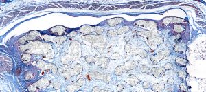
Sarah Just, a doctoral student at the Technical University of Munich (TUM), describes how the M8 digital microscope and slide scanner dramatically changed the workflows and processes of her research in the lab.

Challenges to be solved:
The time it took us to create images of the whole sample was exceptionally long. We used a traditional microscope with a camera on the top that exported the pictures onto a computer. It could only take one image at the magnification of the objective, so for an overview, we took the picture at 10x objective, then changed the objective to a higher magnification for specific areas. Also, to zoom in and out of particular sections, we needed to change objectives and repeat the process. Some of the research we conduct is to compare the sterile mice’s intestines and mice’s intestines with bacteria. We do immunohistochemical staining and then count how many cells exist in a specific area or measure the length of the colon, villi, and ileum. This is also not so convenient to do with analog measuring methods. This is also not so convenient to do with analog measuring methods.
Stay Ahead with Insights from Precipoint!
Welcome to our newsletter! Be the first to know about our latest products, services, webinars, and happenings in PreciPoint. Don't miss out on this opportunity to stay informed. Subscribe to our newsletter today!
By clicking “Subscribe”, you agree to our privacy policy.
I like the microscope because it is much easier to handle, and you can standardize the scanning process. With the scan function, no I do not need to sit in front of a microscope forever. The continuous zoom function is also great.”

Our approach and solution:
With the M8, we have an overview of the slide immediately at high-resolution magnification. The digital microscope is much quicker, and we have a better orientation. Now, it is possible to zoom into our area of interest and to know exactly where we are on the slide. With the overview whole slide image there is no need to switch between pictures during annotations. Previously, we had to take measurements separately from each picture of the microscope slides. We scan almost all the slides so that we can work with them later with ViewPoint (PreciPoint’s free viewer software). For annotations, we use the counting mode, length measurer, and area calculations.


