
Synopsis
An intraoperative frozen section examination is requested by a surgeon during an ongoing operation. With the help of its findings, he can decide on further surgical procedures. In cancer surgery, frozen section is of great importance and can significantly reduce follow-up operations. Nevertheless, this intraoperative service is not always available everywhere. That is because, while on the one hand, frozen section diagnostics poses challenges, on the other, the shortage of pathologists limits its availability. Digital microscopes open up new possibilities for cost-efficient and location-independent collaboration among medical professionals. This can increase the availability of intraoperative services and prove overall cancer treatment for patients.
Real-time Pathology Guidance During Surgery
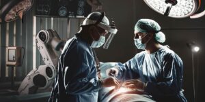
Intraoperative consultation, often also referred to as frozen section, is a pathology service and an important part of surgical pathology. The surgeon requests a frozen section (cryosection) during an ongoing operation. He removes a surgical specimen and sends it to pathology for microscopic frozen section examination. Meanwhile, the operation is briefly interrupted, but the patient remains under anesthesia.
It is possible to perform the intraoperative frozen section within a maximum of 20 minutes after delivery of the specimen to the pathology laboratory. For this purpose, the specimen is first prepared according to the frozen section protocol. The tissue sample is snap-frozen, cut into thin slices, and stained to better visualize possible pathological changes. Finally, the pathologist evaluates the slide under the microscope and communicates his findings to the operating surgeon.
Why Are Frozen Sections Performed?
The frozen section examination provides the surgeon with important information during the operation to decide on its further course, e.g., whether a tumor is benign or malignant. If the tumor is malignant, the surgical incision margins are also examined to check whether cancer has been removed from healthy tissues. Intraoperative frozen section diagnostics can significantly reduce the rate of follow-up surgeries, thus, optimizing overall surgical patient management.
What Are the Challenges of Intraoperative Consultations?
Intraoperative consultations are still not a common practice in surgical cancer therapy. This is because frozen section diagnostics come with some challenges:
Intraoperative Consultations Occur Under High Time Pressure
Within 15 to 20 minutes from receipt of the specimen in the laboratory (depending on country regulations), the pathologist must have transmitted his findings to the operating physician. Within this time frame, macroscopic examination, specimen preparation, and microscopic evaluation must be performed.
Shortage of Pathologists Limits Availability of Intraoperative Services
Many hospitals do not have an in-house pathology department or not the right specialist. Specimens must be sent to outside laboratories, or a pathologist comes to the hospital to perform the consultation. Frozen section examination then involves transportation and travel time. During this time, the operating room is occupied, and the required personnel is blocked without being able to perform other tasks. This also increases the total cost of the operation.
Frozen Section Specimens Are of Lower Quality Than Permanent Specimens
Frozen section specimens obtained by sectioning frozen tissue blocks are generally not as flat as permanent specimens obtained from FFPE blocks. Frozen sections are therefore more difficult to evaluate under the microscope. They require experienced pathologists as well as longer scan times for digitization. Whole slide image (WSI) scans are practically unsuitable for performing frozen section examinations, as the time required to digitize several frozen section specimens with difficult specimen topography is too long.
Why Are Digital Microscopes Particularly Well Suited to be Used for Intraoperative Consultations?
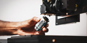
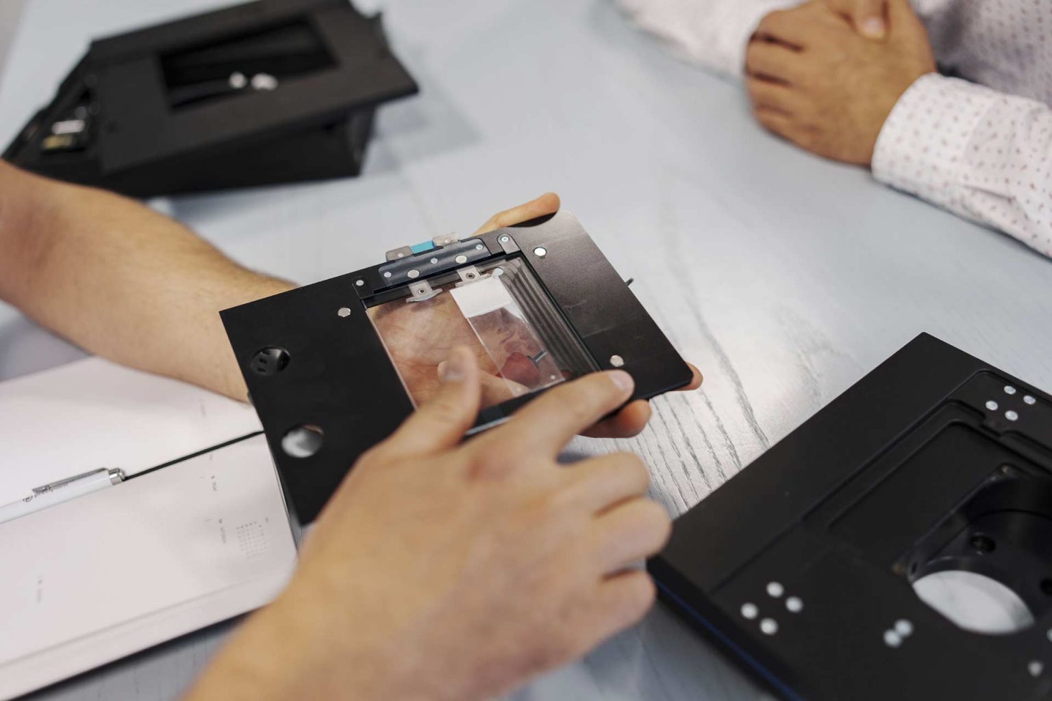
Digital microscopes such as the iO:M8 by PreciPoint have significant advantages, especially for time-critical frozen sections or other intraoperative procedures.
Fast Digitization of Slides
By using digital microscopes, frozen section specimens are quickly available on screen for microscopic evaluation. Pathologists can thus evaluate microscopic specimens in real-time on the computer without having to wait for a WSI scan.
Quick Access to Second Opinions
Not all frozen section examinations are simple cases. They often require special pathological expertise. However, due to high time pressure, there is rarely enough time to obtain a second opinion. Digitized samples visualized on a monitor can be easily and quickly shared with specialists elsewhere via streaming software to obtain expert opinions.
Reducing the Care Gap
Working with digital microscopes and streaming software makes it possible to call in specialists. Furthermore, pathology services can be provided to more healthcare facilities overall without incurring long transportation and travel times.
Using digital microscopes, pathologists can evaluate frozen section specimens in real-time on a computer without having to wait for a WSI scan. In addition, streaming software allows surgeons and pathologists to collaborate. This makes transportation and travel times obsolete. Digital microscopes thus speed up the process of intraoperative consultation and increase its availability. This improves treatment for cancer patients – and that is at an even lower cost for hospital management.
Conclusion
Intraoperative frozen section examinations are performed under high time pressure: The entire process up to delivering the findings may take a maximum of up to 20 minutes. However, frozen section specimens require specific microscopy tools due to their lower sample quality. Also, the shortage of pathologists requires that additional transport or travel times are accepted. These challenges currently limit the availability of intraoperative consultations. Digital microscopes like the iO:M8 offer benefits, particularly in time-sensitive scenarios such as frozen sections during intraoperative procedures. It allows for rapid on-screen access to frozen section specimens, enabling pathologists to instantly assess microscopic details on a computer screen, eliminating the need to wait for a Whole Slide Imaging (WSI) scan. Furthermore, digitized samples displayed on a monitor can be effortlessly and swiftly shared with specialists located remotely through streaming software, facilitating prompt expert consultations.