
Every year, on February 4, World Cancer Day marks an opportunity to raise awareness of this serious health problem on a global level. It’s the initiative of the Union for International Cancer Control (UICC), the largest and oldest international cancer organization. Cancer is the second most common cause of death around the world, and 70% of deaths happen in low-income countries. As evident, constructive actions on all levels, from medical to social and political, are needed. Although the UICC’s activities this year have a social emphasis, the medical aspect cannot be neglected. This illness has a staggering death rate. Every year, approximately 9.6 million people die from this disease.
The Importance of Digital Microscopes in Fighting Cancer
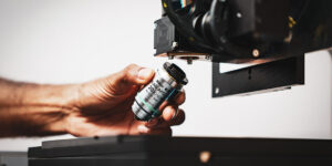
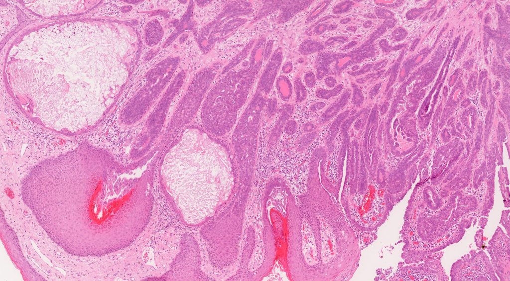
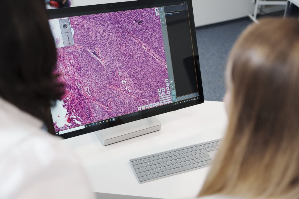
A cancer diagnosis is one of the most important aspects of the treatment and management of the disease. The role of technology in this process is increasingly important, as it offers a lesser scope of error. One example of particularly useful technology in establishing an accurate cancer diagnosis is a digital microscope. Microscope slides are digitized, and pathologists can view high-precision images directly on a computer screen. As a result, the diagnosis not only becomes more accurate but also more consistent.
Pathologists and other experts can take samples and analyze them very quickly, easily, and precisely. Depending on the case, a medical expert can use a digital microscope for research on cancer cells in the sample or determine the diagnosis. If the case is complex and there is a need for collaboration with colleagues from other workstations, an oncologist, pathologist, or other medical specialists can easily share and analyze high-resolution images and videos with each other. They can examine and study the images in every phase of the research and diagnostics.
Stay Ahead with Insights from Precipoint!
Welcome to our newsletter! Be the first to know about our latest products, services, webinars, and happenings in PreciPoint. Don't miss out on this opportunity to stay informed. Subscribe to our newsletter today!
By clicking “Subscribe”, you agree to our privacy policy.
Remote Collaboration and Access with Digital Microscopes
Digital microscopes make remote consulting also possible. Diverse members of the team can communicate remotely and provide their valuable insights. Remote access to the images is especially useful if some team members are working remotely. In that way, a digital microscope breaks the boundaries of a work environment where medical resources and knowledge are limited due to geographical and physical limitations.
Integration of AI for Enhanced Diagnosis
If a digital microscope is integrated with other emerging technologies such as artificial intelligence (AI) or machine learning, the detection of illness and subsequent diagnosis can be more efficient. The accuracy of a cancer diagnosis can be enhanced if AI algorithms are trained to automatically identify and classify cancer cells.
Study on Digital Microscopes and Diagnosis Time
To vouch for the efficiency of digital microscopes, it’s interesting to refer to one study that examined the relationship between digital microscopes and diagnosis time. “Digital pathology has the potential to provide significant benefits in diagnostic pathology,” according to Dr. Emily Clarke et al.’s research at the University of Leeds.
Quicker Diagnosis with Digital Microscopes
The researchers wanted to prove that presenting digital slides for simultaneous viewing of multiple sections of tissue for comparison, as in those with immunohistochemical panels, would allow pathologists to review cases more quickly. Sixteen histopathologists used the digital microscope to review three liver biopsy cases, including an immunohistochemical panel, and three different liver biopsy cases, including an immunohistochemical panel, using the light microscope. They recorded the time of the diagnosis.
Time Efficiency and Accuracy with Digital Microscopes
Those who used a digital microscope needed 30% less time for diagnosis than those who used a light microscope. They reviewed immunohistochemical slides more quickly without incurring any major diagnostic errors. As digital pathology becomes more integrated into the routine, the researchers expected that other tasks would also become more time-efficient.
Fundraisings: An Option to Tackle Financial Challenges
Diverse medical institutions are aware that this kind of equipment has a significant cost, as does the special training of individuals. Every digital microscope needs to be dependable. That means high-quality results that cannot be achieved if, for example, digital images are of low quality. Problems like this can be solved by investing in high-quality cameras, lighting equipment, and even image stabilization and noise reduction software to improve image capture quality. Each of these elements is costly. Fundraising is one of the best solutions to tackle these challenges. Medical organizations, governments, and NGOs often organize luncheons, dinners, and similar events to raise funds. Efficient prevention is impossible without strong collaboration that makes getting digital tools easier.

Although there are some limitations, mostly regarding finances, the future of digital pathology is promising. There is a notable potential to improve the accuracy and consistency of the diagnoses using quick, efficient, and precise methods and tools. With an accurate diagnosis, medical specialists can manage the disease more efficiently, and patients can receive the best treatment possible.