Traditionally, teaching content for students throughout their education is delivered via textbooks, lectures and the use of a conventional light microscope.
However, digitally supported teaching concepts allow many things to be done more efficiently and also better. In this short article, learn how virtual microscopy is being evaluated and embraced by both students and faculty.
A study was conducted at the Medical College of Wisconsin to investigate the learning success with virtual microscopy compared to conventional light microscopy. Other universities have also digitized their courses and published the results.
Used virtual microscopy for first-year medical students. Students use the new technology for their courses in histology, cell and tissue biology, and integrated medical neuroscience.

Why are universities switching to virtual microscopy?
The switch was prompted by maintenance difficulties and the cost of replacing the college’s microscopes and slides, and primarily by a desire to make learning easier and more efficient for large classes (over 200 students) of first-year medical students.
After participating in the courses, students and faculty were surveyed about their experience with virtual microscopy. Exam results, ease of use, effectiveness for learning and teaching, personal preference over light microscopy, and other aspects were examined.
Stay Ahead with Insights from Precipoint!
Welcome to our newsletter! Be the first to know about our latest products, services, webinars, and happenings in PreciPoint. Don't miss out on this opportunity to stay informed. Subscribe to our newsletter today!
By clicking “Subscribe”, you agree to our privacy policy.
Do students prefer virtual microscopy over light microscopy?
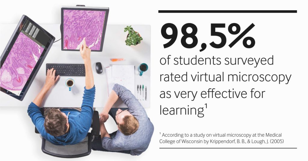
Students were asked to subjectively rate the overall effectiveness of the virtual microscope for learning. In this survey, 98.5% rated the effectiveness of the virtual microscope for learning as excellent or good [1].
There was also room for personal comments on the surveys, which were overly enthusiastic. One student provided the following feedback in the survey:
"[The virtual microscope] eliminated a lot of frustration I have had in the past with other microscopes.... Easier to ask questions and discuss slides with other people since we could all look at the same thing."[1]
Krippendorf, B. B., & Lough, J. (2005). Tweet
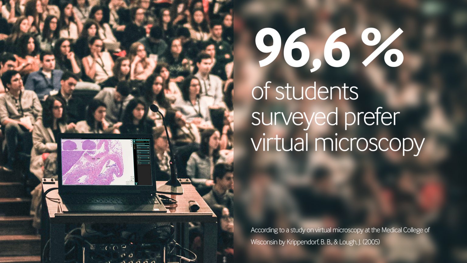
Another question on the survey dealt with whether students preferred learning via the virtual microscope to learning with the light microscope. Again, the results were clearly in favor of the virtual microscope, with 96.6% of first semester students agreeing or strongly agreeing that they preferred learning via the virtual microscope. Of the second-semester students who had been taught using light microscopy in the previous year, 72.8% were still clearly in favor of the virtual microscope [1].
The instructors were also convinced of the use of the digital method. Out of the 12 respondents, 10 showed a strong preference for virtual microscopy. One instructor had no preference, and only one of the 12 votes went to light microscopy [1].
The Medical College of Wisconsin is not the only university to host its courses using virtual microscopy. For example, the University of Belfast started a similar project in 2006 and equipped their histology courses with virtual microscopy. As in Wisconsin, second-year medical students readily embraced the technology and could envision continuing to work with digital specimens. Of the 136 survey participants, 88% said they preferred virtual microscopy. Eleven percent, however, felt that microscopy skills would suffer in virtual-only classes, which they believe are important. Nearly all students (99%) found the digital specimens easy to navigate and 93% found the image quality at least as good as that of a regular microscope. In addition, tutors noted that students were more interested in the slides [2].
Almost all faculty members teaching histology laboratories were convinced of the switch from light to virtual microscopy. A sizable proportion felt that virtual microscopy made teaching much easier and more efficient and made group communication much simpler. The result is not surprising now that digital devices have become commonplace in our everyday lives. This effect is likely to intensify over time as younger generations grow up with the increasingly digital technology.
[1] Krippendorf, B. B., & Lough, J. (2005). Complete and rapid switch from light microscopy to virtual microscopy for teaching medical histology. The Anatomical Record Part B: The New Anatomist: An Official Publication of the American Association of Anatomists, 285(1), 19-25.
[2] Hamilton, P. W., Wang, Y., & McCullough, S. J. (2012). Virtual microscopy and digital pathology in training and education. Apmis, 120(4), 305-315.


